The movement nobody looks at
The Movement pillar describes nine loading patterns — squat, hip hinge, pushes, pulls, lunge, loaded carry, rotation. It was missing the one everybody performs thousands of times a day.
This is not an inventory detail. Eight pages in this corpus point to walking to explain what a muscle is for — tibialis anterior, the calves, gluteus medius, the iliopsoas and others — without any page describing the movement itself.
Gait cycle
The interval between two successive contacts of the same foot with the ground. It divides into a stance phase, where the foot is on the ground, and a swing phase, where it travels freely.
stride
What defines walking
A single rule separates walking from running, and it is not speed.
Certainty level · Established
In walking, at least one foot is always in contact with the ground, and there are two periods where both feet touch the ground at once.
This is the mechanical definition used in gait analysis: the stance phase occupies around three fifths of the cycle and swing the remaining two fifths, which leaves an overlap at each change of foot. Those proportions tighten as pace increases, and double support disappears at the transition to running.
Perry J, Burnfield JM (2010) · Novacheck TF (1998)
That rule has a direct consequence: walking has no airborne phase. The body never falls back down, and the forces it takes stay close to its own weight.
The phases of a step
One cycle, from one heel contact to the next
- 1Initial contact0 %The heel touches the ground ahead of the body. The foot is about to go flat.
- 2Loading response0–12 %Weight transfers onto this leg. Tibialis anterior brakes the descent of the forefoot, the quadriceps absorbs the slight knee flexion.
- 3Mid-stance12–50 %The body passes over the planted foot. This is single-leg stance: the other leg is in the air.
- 4Terminal stance50–62 %The heel lifts, the calf propels. The hip is at maximum extension.
- 5Pre-swing and swing62–100 %The toe leaves the ground, the leg travels forward, and the hamstrings brake its advance before the next contact.
The problem nobody notices
At every step, for more than a third of the cycle, the whole weight of the body rests on one leg. The pelvis should then tip towards the unsupported side, like a plank set on a single pillar.
Certainty level · Established
During single-leg stance, the hip abductors on the standing side produce the force that keeps the pelvis from dropping on the opposite side.
The mechanism is established by kinesiology and confirmed by instrumented gait analysis, which measures pelvic tilt and muscle activity through the cycle. How much residual drop remains varies between people and with speed.
Perry J, Burnfield JM (2010) · Neumann DA (2016)
This is the reason gluteus medius exists, and the only occasion where that deep muscle becomes noticeable in ordinary life. Tensor fasciae latae and the hip adductors take part in the same sideways control.
Braking more than propelling
The movement looks propulsive, but most of the muscles involved work in the eccentric regime — they hold back.
Who does what, and in which regime
Muscle
Moment in the cycle
Regime
What it does
Tibialis anterior
After heel contact
Eccentric
Brakes the forefoot landing
Quadriceps
Loading response
Eccentric
Absorbs the knee flexion
Calves
Terminal stance
Concentric
Lift the heel and propel
Hamstrings
End of swing
Eccentric
Brake the advancing leg
Gluteus medius
All of single-leg stance
Isometric
Keeps the pelvis level
Only one of those five groups actually produces the movement. The others organise it, brake it or keep it from deviating.
Why walking costs so little
Walking is remarkably economical, and the reason is mechanical rather than muscular.
At every step the body rises slightly as it passes over the planted foot, then comes back down. It thereby trades positional energy for movement energy, like an inverted pendulum: what is gained in height at mid-stance is returned as speed at the end. The muscle only has to make up the losses.
Key point
What changes when running
That exchange works as long as a foot stays on the ground. As soon as an airborne phase appears, the mechanism switches: the body no longer swings, it bounces, and energy is no longer exchanged but stored in the tendons. The running page describes that switch.
Seeing the muscles involved
The deep and medial plane
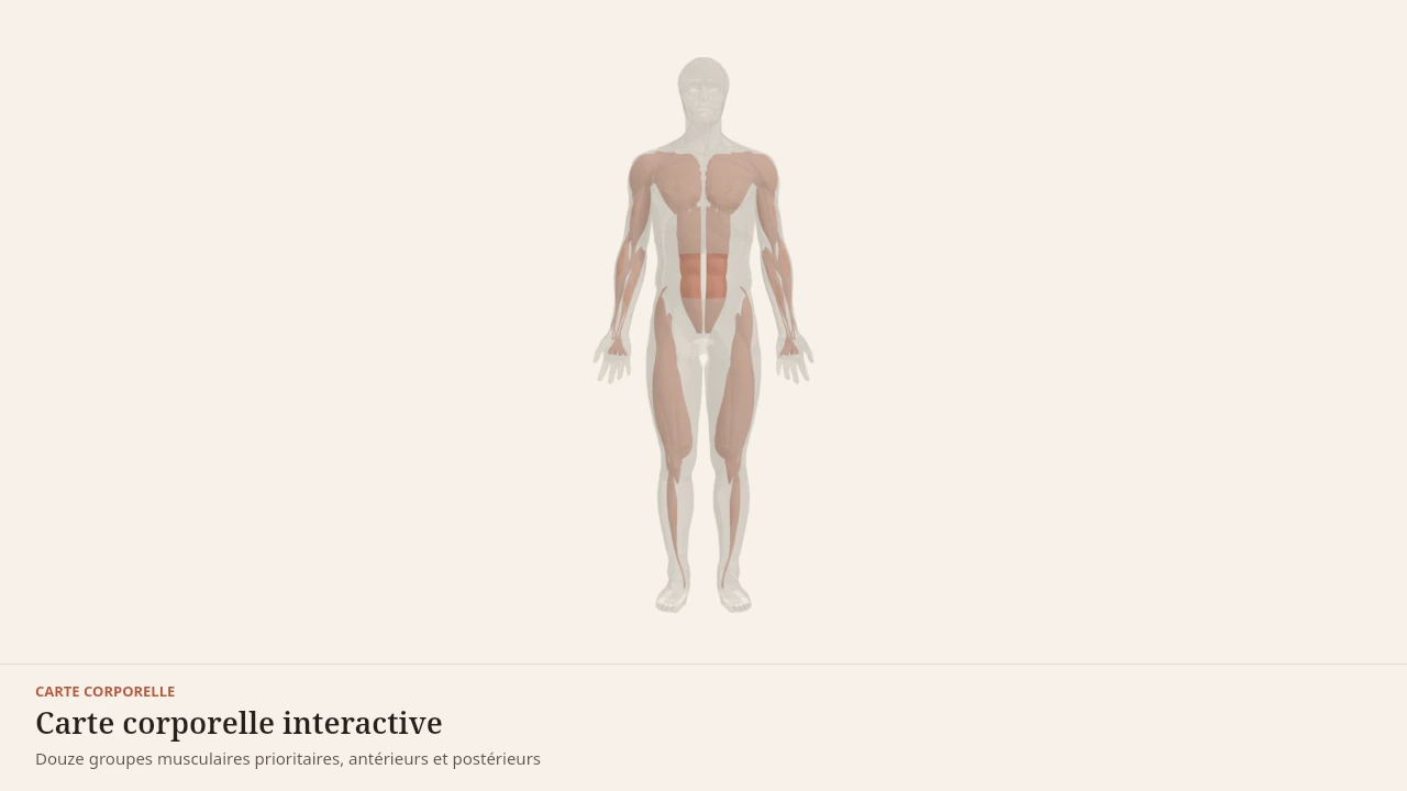
The scene shows the muscles walking calls on without any outside view revealing them: gluteus medius, the iliopsoas and the adductors. Without the 3D, the logic is enough: the muscles that hold the pelvis through a step are not the ones you can see.
Current step
Deep and medial muscles
Beneath the visible groups work muscles no silhouette shows. The rotator cuff holds the head of the humerus against the shoulder blade, serratus anterior keeps the shoulder blade flat on the ribcage, the iliopsoas links the lumbar spine to the femur, and the adductors fill the inner thigh. Deeper still, the diaphragm closes the trunk as a dome, inside the ribs. They orient and stabilise more than they move.
Scene description
A three-dimensional diagram of a standing human body, arms by the sides, built from simplified paper-coloured volumes. Twelve priority muscle groups are laid over this outline in terracotta: at the front, pectoralis major, the deltoid, biceps brachii, the abdominal wall and the quadriceps; at the back, the trapezius, latissimus dorsi, the erector spinae, triceps brachii, the glutes, the hamstrings and the calves. A simplified skeleton — skull, spine, ribcage, pelvis, humerus, femur and tibia — can be shown beneath the outline to reveal the bony levers these muscles pull on. Each group is selectable and opens its own reference page. This is a teaching diagram with stylised volumes, not an exact anatomical reconstruction.
Visible structures
- Pectoralis majorA thick sheet running from the clavicle, the sternum and the upper costal cartilages to the lateral lip of the bicipital groove of the humerus. It draws the arm towards the midline and forwards, and drives horizontal pushing.
- DeltoidsThree heads — anterior, lateral and posterior — running from the clavicle, the acromion and the spine of the scapula to the deltoid tuberosity of the humerus. Together they raise the arm; separately they carry it forwards, out to the side or backwards.
- Biceps brachiiTwo heads arising from the scapula — the supraglenoid tubercle and the coracoid process — joining on the radial tuberosity. It flexes the elbow and supinates the forearm, and contributes to every pulling movement.
- Abdominal wallRectus abdominis, the external and internal obliques and transversus, running from the ribs and sternum to the pubis and iliac crest. They flex and rotate the trunk and, above all, stabilise the spine by regulating intra-abdominal pressure.
- QuadricepsFour heads — rectus femoris, vastus lateralis, vastus medialis and vastus intermedius — joining through the quadriceps tendon onto the patella and then the tibial tuberosity. They extend the knee, and rectus femoris also flexes the hip.
- TrapeziusA diamond-shaped muscle spanning from the occiput and the cervical and thoracic spinous processes to the clavicle, the acromion and the spine of the scapula. Its upper, middle and lower portions elevate, retract and depress the scapula.
- Latissimus dorsiA broad fan arising from the lower thoracic vertebrae, the thoracolumbar fascia, the iliac crest and the lowest ribs, converging on the bicipital groove of the humerus. It pulls the arm down and back, and drives vertical pulling.
- Erector spinaeMuscular columns — iliocostalis, longissimus and spinalis — running from the sacrum and iliac crest to the ribs, the vertebrae and the skull, on either side of the spinous processes. They extend the spine and control the trunk as it lowers.
- Triceps brachiiThree heads — long, lateral and medial — arising from the scapula and the back of the humerus and joining on the olecranon of the ulna. It extends the elbow, and its long head also assists shoulder extension.
- GlutesGluteus maximus, medius and minimus, arising from the iliac wing and the sacrum and inserting on the femur and the iliotibial tract. They extend, abduct and rotate the hip, and drive the pelvis upright in the hinge and the squat.
- HamstringsBiceps femoris, semitendinosus and semimembranosus, running from the ischial tuberosity to the tibia and fibula. They extend the hip and flex the knee, and control the trunk as it lowers in the hip hinge.
- CalvesThe gastrocnemius and soleus, joining through the Achilles tendon onto the calcaneus. They plantarflex the ankle, and the gastrocnemius, which crosses the knee, also assists knee flexion.
- Rotator cuffFour short muscles running from the shoulder blade to the top of the humerus. Their tendons merge into a sheet that holds the head of the bone against its socket while the deltoid moves the arm.
- Serratus anteriorA sheet hooked onto the upper ribs by triangular slips, passing under the shoulder blade and attaching to its inner border. It holds the blade against the ribcage and rotates it.
- IliopsoasThe only muscle linking the lumbar spine directly to the femur. It descends from the vertebrae and the iliac fossa, curves around the rim of the pelvis and attaches to the lesser trochanter.
- Hip adductorsFive muscles running from the pubis to the back of the femur in overlapping layers. They draw the thigh towards the midline and control the pelvis in single-leg stance.
- Forearm musclesMasses gathered near the elbow, continued by long tendons to the wrist and fingers. They drive the hand from a distance and carry grip strength.
- Tibialis anteriorA muscle pressed against the outer surface of the tibia, its tendon swinging towards the inside of the foot. It lifts the toes on every step and brakes the forefoot after heel strike.
- DiaphragmA muscular dome running from the lower border of the ribs, the sternum and the lumbar vertebrae to a central tendon. As it contracts it descends, the chest gains volume and air enters: it is the engine of quiet breathing, and the ceiling of the abdominal cavity.
Guided steps
- 1/5The front of the body. Pectoralis major covers the upper chest, the deltoid caps the shoulder, the biceps fills the front of the arm, the abdominal wall links the ribs to the pelvis, and the quadriceps fills the front of the thigh. Further out on the limbs, the forearm muscles drive the wrist and hand, and tibialis anterior lifts the foot.
- 2/5The back of the body. The trapezius links the neck to the shoulder blades, the latissimus dorsi fans down to the pelvis, the erector spinae flank the spine, and the triceps fills the back of the arm. Glutes, hamstrings and calves form the posterior chain from hip to ankle.
- 3/5Beneath the visible groups work muscles no silhouette shows. The rotator cuff holds the head of the humerus against the shoulder blade, serratus anterior keeps the shoulder blade flat on the ribcage, the iliopsoas links the lumbar spine to the femur, and the adductors fill the inner thigh. Deeper still, the diaphragm closes the trunk as a dome, inside the ribs. They orient and stabilise more than they move.
- 4/5In a push, force starts at the ground, travels through the quadriceps and the abdominal wall, and then pectoralis major, the anterior deltoid and the triceps drive the load away from the trunk. The skeleton acts as the lever: no group works alone, and the quality of the movement comes from their sequencing.
- 5/5In a pull, the latissimus dorsi and trapezius bring the arm and shoulder blade towards the trunk while the biceps flexes the elbow. Glutes, hamstrings and erector spinae hold the pelvis and spine so that force transfers without the trunk collapsing.
Model licence · Z-Anatomy et BodyParts3D — CC-BY-SA 4.0
What the model does not show
Limit
A scene cannot walk
Body Lab's anatomical scenes are fixed meshes, derived from sources carrying no animation skeleton. Rotating a bone there would tear away the muscle attached to it. No scene can therefore play a step.
This page consequently describes a movement the model only locates. The sequence in time is carried by the text and the timeline, not by the 3D.
Common misunderstandings
Caution
Three received ideas
That walking is a movement of the legs. The pelvis, the trunk and the arms take part continuously, and the most constant work in the cycle is sideways holding.
That speed distinguishes walking from running. What distinguishes them is the presence or absence of an instant with no support. Fast walking is still walking.
That one step is symmetrical and identical to the next. Instrumented analysis shows differences between the two sides in most people. Interpreting them is clinical work, not page-reading.
Walking extends the lunge, which shares its narrow-base problem, and underpins the loaded carry, which is nothing but loaded walking. On the structural side it calls on the calves, tibialis anterior, gluteus medius, the hamstrings and the iliopsoas.
Sources
Main sources
- Perry J, Burnfield JM (2010). Gait Analysis: Normal and Pathological Function, 2nd edition. SLACK Incorporated.https://www.ncbi.nlm.nih.gov/nlmcatalog/101519549
- Neumann DA (2016). Kinesiology of the Musculoskeletal System: Foundations for Rehabilitation, 3rd edition. Elsevier.https://shop.elsevier.com/books/kinesiology-of-the-musculoskeletal-system/neumann/978-0-323-28753-1
- Novacheck TF (1998). The biomechanics of running. Gait & Posture.DOI : 10.1016/s0966-6362(97)00038-6
Put it into practice in Shapier
Walking with a load
Training the loaded carry, which is nothing but loaded walking, is done in Shapier; Body Lab only explains why walking is work of control as much as of propulsion.
Walking with a load
Body Lab explains; Shapier lets you act and track.
Check my understanding
What mechanically distinguishes walking from running?
In walking, at least one foot is always in contact with the ground, with two periods of double support.
The speed of travel.
The time spent on a single leg.
Which contraction regime dominates muscular work during walking?
The eccentric regime: most muscles brake or hold the movement back.
The concentric regime, because walking is a propulsive movement.
The isometric regime, because the body stays upright.
What keeps the pelvis from dropping on the unsupported side during single-leg stance?
The hip abductors on the standing side, including the gluteus medius.
The trunk and arms, which rebalance the body.
Nothing: the pelvis tips freely at every step.
Choose an answer
Read next
- RunningWhat separates running from walking is not speed but an instant with no support. That instant changes everything: the forces taken, the role of the tendons and the way energy circulates.With a 3D scene
- Gluteus mediusIt pushes nothing forward. Its work is sideways: stopping the pelvis dropping on the unsupported side, on every step — roughly half the time you spend walking.With a 3D scene
- Contraction regimesA muscle can produce force while shortening, while lengthening, or without changing length. It produces the most while lengthening — which is why you can lower a load you cannot lift.With a 3D scene
Available offline
Offline download is available in the mobile app.
Trust and method
Author
Editorial review
Thanh Chau
Published on
August 2, 2026
Reviewed on August 2, 2026
Next review due August 2, 2027
Limits of this page
- The proportions of the cycle described here hold for ordinary-pace walking on level ground in healthy adults. They change with speed, slope, terrain and age.
- Body Lab's scenes are fixed meshes with no animation skeleton: none of them can show a step. This page describes a movement the model does not play.
- Gait analysis is a clinical tool. This page describes a general functioning and allows no assessment of any particular way of walking.
- A limp, pain appearing while walking or a loss of balance belongs to a health professional.
Educational content. Body Lab does not diagnose and does not replace professional advice.
How we work