A muscle that touches no bone below
Tensor fasciae latae is short — around fifteen centimetres — and sits at the front of the outer hip. What sets it apart is its insertion: it attaches not to a bone, but to a fibrous band.
That band, the iliotibial band, runs down the whole outer face of the thigh and attaches to the tibia, below the knee. The muscle therefore pulls on a cord, and the cord carries the pull far beyond it.
Iliotibial band
The fibrous thickening of the outer face of the fascia lata, the connective wrapping of the thigh. It runs from the iliac crest to the tibia and receives the insertions of tensor fasciae latae and part of gluteus maximus.
iliotibial tract
Origin
It arises from the front part of the iliac crest and the anterior superior iliac spine, the bony prominence you can feel at the front of the hip.
Insertion
After a few centimetres its fibres run into the iliotibial band, at the junction of the upper third and lower two-thirds of the thigh.
Nerve supply and palpable landmarks
The nerve supply comes from the superior gluteal nerve, from the L4 to S1 roots — the same as gluteus medius. The muscle can be felt at the front of the hip, just below the iliac spine, and hardens when you lift the leg sideways.
What tensor fasciae latae does
It acts on two joints without directly touching either from below.
- Hip abduction: it takes the thigh outwards, alongside gluteus medius.
- Hip flexion: its forward origin gives it a flexing component.
- Medial rotation: it turns the thigh inwards.
- Action on the knee: through the band, it takes part in sideways knee stability, with a contribution that changes with the angle of flexion.
Certainty level · Established
Tensor fasciae latae transmits its pull to the tibia through the iliotibial band, with no bony insertion of its own at the knee.
Descriptive anatomy and kinesiology describe the same arrangement. The exact share of the band in sideways knee stability varies with the angle of flexion and with measurement method.
Standring S (2020) · Neumann DA (2016)
The first fascia visible in a scene
The fascia page explains that this tissue appears in no model, for want of a separate mesh in the sources used. The iliotibial band is the exception: it is thick and distinct enough to exist as a separate part.
It is therefore, in the whole corpus, the only fascial structure that can be seen. It illustrates well what the fascia page describes in general: a wrapping that is not passive, that muscles attach to and through which force is transmitted.
The movements it takes part in
It works in the lunge and in every reduced-base position, where sideways control of the pelvis becomes a constraint. It also works in walking and running, on every single-leg stance, with gluteus medius.
Synergists
- Gluteus medius shares its nerve supply and its abduction role.
- The upper fibres of gluteus maximus also end on the band, from behind.
- The iliopsoas accompanies its flexing component.
Antagonists
- The hip adductors bring the thigh back towards the midline.
- The lateral hip rotators oppose its medial rotation component.
Seeing the tensor and its band
Tensor fasciae latae and its band
The scene shows the muscle at the front of the hip and the fibrous band running down from it to the tibia. Seen from the front, the band is almost edge-on: it appears as a line along the outer border of the thigh rather than as a sheet. The two are also merged into a single volume, the boundary between fleshy belly and band not being drawn. Without the 3D, the picture fits in one sentence: a short muscle at the top, a long strap below.
Current step
Anterior view
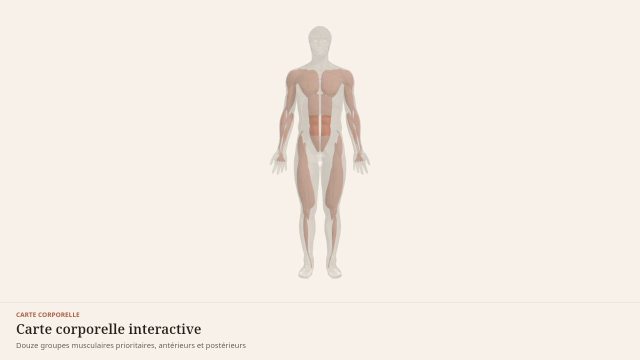
The front of the body. Pectoralis major covers the upper chest, the deltoid caps the shoulder, the biceps fills the front of the arm, the abdominal wall links the ribs to the pelvis, and the quadriceps fills the front of the thigh. Further out on the limbs, the forearm muscles drive the wrist and hand, and tibialis anterior lifts the foot.
Scene description
A three-dimensional diagram of a standing human body, arms by the sides, built from simplified paper-coloured volumes. Twelve priority muscle groups are laid over this outline in terracotta: at the front, pectoralis major, the deltoid, biceps brachii, the abdominal wall and the quadriceps; at the back, the trapezius, latissimus dorsi, the erector spinae, triceps brachii, the glutes, the hamstrings and the calves. A simplified skeleton — skull, spine, ribcage, pelvis, humerus, femur and tibia — can be shown beneath the outline to reveal the bony levers these muscles pull on. Each group is selectable and opens its own reference page. This is a teaching diagram with stylised volumes, not an exact anatomical reconstruction.
Visible structures
- Pectoralis majorA thick sheet running from the clavicle, the sternum and the upper costal cartilages to the lateral lip of the bicipital groove of the humerus. It draws the arm towards the midline and forwards, and drives horizontal pushing.
- DeltoidsThree heads — anterior, lateral and posterior — running from the clavicle, the acromion and the spine of the scapula to the deltoid tuberosity of the humerus. Together they raise the arm; separately they carry it forwards, out to the side or backwards.
- Biceps brachiiTwo heads arising from the scapula — the supraglenoid tubercle and the coracoid process — joining on the radial tuberosity. It flexes the elbow and supinates the forearm, and contributes to every pulling movement.
- Abdominal wallRectus abdominis, the external and internal obliques and transversus, running from the ribs and sternum to the pubis and iliac crest. They flex and rotate the trunk and, above all, stabilise the spine by regulating intra-abdominal pressure.
- QuadricepsFour heads — rectus femoris, vastus lateralis, vastus medialis and vastus intermedius — joining through the quadriceps tendon onto the patella and then the tibial tuberosity. They extend the knee, and rectus femoris also flexes the hip.
- TrapeziusA diamond-shaped muscle spanning from the occiput and the cervical and thoracic spinous processes to the clavicle, the acromion and the spine of the scapula. Its upper, middle and lower portions elevate, retract and depress the scapula.
- Latissimus dorsiA broad fan arising from the lower thoracic vertebrae, the thoracolumbar fascia, the iliac crest and the lowest ribs, converging on the bicipital groove of the humerus. It pulls the arm down and back, and drives vertical pulling.
- Erector spinaeMuscular columns — iliocostalis, longissimus and spinalis — running from the sacrum and iliac crest to the ribs, the vertebrae and the skull, on either side of the spinous processes. They extend the spine and control the trunk as it lowers.
- Triceps brachiiThree heads — long, lateral and medial — arising from the scapula and the back of the humerus and joining on the olecranon of the ulna. It extends the elbow, and its long head also assists shoulder extension.
- GlutesGluteus maximus, medius and minimus, arising from the iliac wing and the sacrum and inserting on the femur and the iliotibial tract. They extend, abduct and rotate the hip, and drive the pelvis upright in the hinge and the squat.
- HamstringsBiceps femoris, semitendinosus and semimembranosus, running from the ischial tuberosity to the tibia and fibula. They extend the hip and flex the knee, and control the trunk as it lowers in the hip hinge.
- CalvesThe gastrocnemius and soleus, joining through the Achilles tendon onto the calcaneus. They plantarflex the ankle, and the gastrocnemius, which crosses the knee, also assists knee flexion.
- Rotator cuffFour short muscles running from the shoulder blade to the top of the humerus. Their tendons merge into a sheet that holds the head of the bone against its socket while the deltoid moves the arm.
- Serratus anteriorA sheet hooked onto the upper ribs by triangular slips, passing under the shoulder blade and attaching to its inner border. It holds the blade against the ribcage and rotates it.
- IliopsoasThe only muscle linking the lumbar spine directly to the femur. It descends from the vertebrae and the iliac fossa, curves around the rim of the pelvis and attaches to the lesser trochanter.
- Hip adductorsFive muscles running from the pubis to the back of the femur in overlapping layers. They draw the thigh towards the midline and control the pelvis in single-leg stance.
- Forearm musclesMasses gathered near the elbow, continued by long tendons to the wrist and fingers. They drive the hand from a distance and carry grip strength.
- Tibialis anteriorA muscle pressed against the outer surface of the tibia, its tendon swinging towards the inside of the foot. It lifts the toes on every step and brakes the forefoot after heel strike.
- DiaphragmA muscular dome running from the lower border of the ribs, the sternum and the lumbar vertebrae to a central tendon. As it contracts it descends, the chest gains volume and air enters: it is the engine of quiet breathing, and the ceiling of the abdominal cavity.
Guided steps
- 1/5The front of the body. Pectoralis major covers the upper chest, the deltoid caps the shoulder, the biceps fills the front of the arm, the abdominal wall links the ribs to the pelvis, and the quadriceps fills the front of the thigh. Further out on the limbs, the forearm muscles drive the wrist and hand, and tibialis anterior lifts the foot.
- 2/5The back of the body. The trapezius links the neck to the shoulder blades, the latissimus dorsi fans down to the pelvis, the erector spinae flank the spine, and the triceps fills the back of the arm. Glutes, hamstrings and calves form the posterior chain from hip to ankle.
- 3/5Beneath the visible groups work muscles no silhouette shows. The rotator cuff holds the head of the humerus against the shoulder blade, serratus anterior keeps the shoulder blade flat on the ribcage, the iliopsoas links the lumbar spine to the femur, and the adductors fill the inner thigh. Deeper still, the diaphragm closes the trunk as a dome, inside the ribs. They orient and stabilise more than they move.
- 4/5In a push, force starts at the ground, travels through the quadriceps and the abdominal wall, and then pectoralis major, the anterior deltoid and the triceps drive the load away from the trunk. The skeleton acts as the lever: no group works alone, and the quality of the movement comes from their sequencing.
- 5/5In a pull, the latissimus dorsi and trapezius bring the arm and shoulder blade towards the trunk while the biceps flexes the elbow. Glutes, hamstrings and erector spinae hold the pelvis and spine so that force transfers without the trunk collapsing.
Model licence · Z-Anatomy et BodyParts3D — CC-BY-SA 4.0
What the model simplifies
Limit
A band without its wrapping
The iliotibial band is the lateral thickening of the fascia lata, which wraps the whole thigh. Only that lateral part exists as a distinct piece in the sources: the rest of the wrapping is not represented.
The muscle and the band are also rendered as a single volume, although the transition from fleshy to fibrous tissue is abrupt.
A subject that calls for care
Caution
What this page does not say
Iliotibial band syndrome is a frequent reason for consultation among runners, and its mechanisms are debated. Describing the anatomy of the structure establishes neither its causes nor grounds for attributing a pain to it.
Body Lab proposes here no exercise, no stretch and no technique. Pain on the outer side of the knee or hip that returns with effort belongs to a health professional.
Sources
Main sources
- Standring S (2020). Gray's Anatomy: The Anatomical Basis of Clinical Practice, 42nd edition. Elsevier.https://shop.elsevier.com/books/grays-anatomy/standring/978-0-7020-7705-0
- Neumann DA (2016). Kinesiology of the Musculoskeletal System: Foundations for Rehabilitation, 3rd edition. Elsevier.https://shop.elsevier.com/books/kinesiology-of-the-musculoskeletal-system/neumann/978-0-323-28753-1
Put it into practice in Shapier
Train tensor fasciae latae with clamshells
Training tensor fasciae latae through hip abduction happens in Shapier with clamshells; Body Lab only explains what this muscle does.
Train tensor fasciae latae with clamshells
Body Lab explains; Shapier lets you act and track.
Check my understanding
What does tensor fasciae latae insert onto?
The iliotibial band, which carries its pull down to the tibia
Directly onto the tibia, below the knee
Onto the femur, at the lower end of the thigh
Why is the iliotibial band the only fascial structure visible in the corpus scenes?
Because it is thick and distinct enough to exist as a separate part
Because fascia appears in every anatomical model
Because it was drawn specially for this page
What should you take away from what the page says about iliotibial band syndrome?
That describing the anatomy of the structure establishes neither its causes nor grounds for attributing a pain to it
That it is caused by a muscle that is too short in runners
That exercises and stretches are enough to treat it
Choose an answer
Read next
- Gluteus mediusIt pushes nothing forward. Its work is sideways: stopping the pelvis dropping on the unsupported side, on every step — roughly half the time you spend walking.With a 3D scene
- FasciaA continuous sheet of connective tissue, real and well described in anatomy. What is commonly said about it goes markedly further than what the literature establishes.Described without a scene
- The kneeTwo poorly matched surfaces, an almost flat plateau under rounded condyles. Menisci, cruciate and collateral ligaments make up for that lack of fit.With a 3D scene
Available offline
Offline download is available in the mobile app.
Trust and method
Author
Editorial review
Thanh Chau
Published on
August 2, 2026
Reviewed on August 2, 2026
Next review due August 2, 2027
Limits of this page
- The muscle and the band are extracted together in the scene: the boundary between the fleshy belly and the fibrous band is not drawn, although it is sharp.
- The fascia lata, of which the band is the lateral thickening, wraps the whole thigh: only its lateral part is represented.
- Iliotibial band syndrome is a clinical subject with debated mechanisms. This page describes an anatomy and establishes neither its causes nor what to do about it.
Educational content. Body Lab does not diagnose and does not replace professional advice.
How we work