Where the triceps brachii is located
The triceps brachii fills the entire back compartment of the upper arm on its own, between the shoulder and the elbow. Its bulk accounts for a large share of arm girth, more than the biceps brachii facing it. Its name comes from its three heads, which start at three different places to join a common tendon.
Origins
- The long head arises from the infraglenoid tubercle of the shoulder blade, just below the socket of the shoulder joint.
- The lateral head arises from the back surface of the humerus, above the groove of the radial nerve.
- The medial head arises from the back surface of the humerus, below that same groove.
Only the long head takes its support from the shoulder blade. The other two start on the arm bone itself, which makes them indifferent to the position of the shoulder.
Insertion
The three heads gather into a thick tendon that attaches to the olecranon, the bony point felt at the back of the elbow. Some of the fibres continue into the fascia of the forearm.
Innervation and palpable landmarks
Innervation comes from the radial nerve, from roots C6 to C8. The olecranon is felt immediately; following the back surface of the arm upward, the mass of the triceps can be traced as far as the armpit.
What the triceps brachii does
The triceps has one main action and one secondary action.
- Elbow extension: the three heads straighten the forearm. It is the only substantial muscle group producing this action.
- Shoulder extension and adduction: the long head, because it also crosses the shoulder joint, draws the arm backward and toward the body.
Biarticular muscle
A muscle that crosses two joints and therefore acts on both. Its total length depends on the combined position of those joints, which changes the force it can produce.
two-joint muscle
Certainty level · Established
The position of the shoulder changes the length of the long head of the triceps, and therefore the tension it can produce at the elbow.
Like any biarticular muscle, the long head is stretched when the shoulder is flexed with the arm above the head, and shortened when the arm is brought down alongside the body. The principle at work is the length–tension relationship, established on isolated preparations and then confirmed on intact muscle. The practical consequence is that an elbow extension does not load the long head in the same way depending on the orientation of the arm. How large that effect is depends on segment lengths and varies from person to person.
Neumann DA (2016)
Seeing the triceps in motion
The triceps in a pushing movement
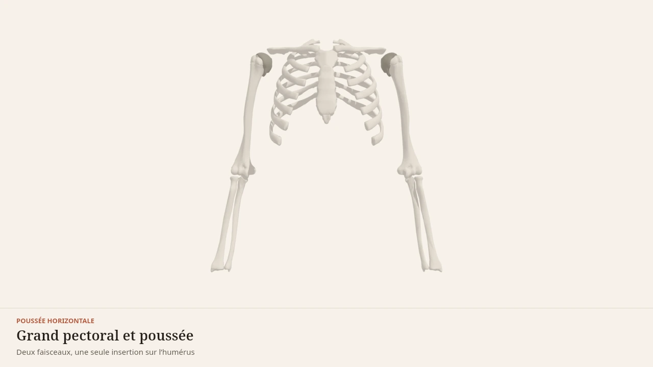
The scene shows the arm during a horizontal push. The triceps shortens as the elbow straightens, while the pectoralis major brings the arms together in front of the chest. Without the 3D, the essential point is that the triceps pulls the olecranon upward, which straightens the forearm.
Current step
The frame of the push
Pectoralis major does not pull into thin air: it links a wide fixed base — clavicle, sternum and costal cartilages — to a single moving point on the humerus. Locating those bony attachments is already enough to understand its line of pull.
Scene description
A three-dimensional diagram of the upper trunk seen from the front, both arms held out horizontally as at the start of a bench press. The ribcage, sternum, both clavicles, both humeri and the forearms are shown in bone tones, and each shoulder joint is marked by a small dark sphere. Over this frame, pectoralis major is laid out in terracotta as two distinct heads: a thin clavicular head under the collarbone, and a much broader sternocostal head running from the sternum to the humerus, both converging on the same insertion point. The anterior deltoid caps the front of the shoulder and triceps brachii runs along the back of the arm. An animation brings the humeri in front of the chest in horizontal adduction while the belly of pectoralis major thickens. The volumes are deliberately simplified: this is a teaching diagram, not an exact anatomical reconstruction.
Visible structures
- Clavicular headThe thin upper portion of pectoralis major, arising from the medial third of the front of the clavicle. Its fibres run down to the humerus, carrying the arm forwards and upwards, and it dominates in incline pushing.
- Sternocostal headThe broad lower portion, arising from the lateral border of the sternum and the cartilages of the first six ribs. Its fibres run obliquely up to the bicipital groove of the humerus, bringing the arm forwards and downwards.
- Humeral insertionBoth heads converge into a flat tendon attached to the lateral lip of the bicipital groove of the humerus. The whole origin — clavicle, sternum and ribs — therefore pulls on this single point, which is what makes the line of pull so directional.
- Anterior deltoidThe front head of the deltoid, arising from the lateral third of the clavicle and inserting on the deltoid tuberosity. It flexes the shoulder and carries the arm forwards, working alongside pectoralis major in every push.
- Triceps brachiiThree heads joining on the olecranon of the ulna. It extends the elbow and finishes the push: without it the arm closes towards the trunk but the load never actually travels away.
Guided steps
- 1/5Pectoralis major does not pull into thin air: it links a wide fixed base — clavicle, sternum and costal cartilages — to a single moving point on the humerus. Locating those bony attachments is already enough to understand its line of pull.
- 2/5The upper portion arises from the medial third of the clavicle. Its fibres run obliquely downwards to the humerus, which gives them a flexing action: they carry the arm forwards and upwards, and dominate in incline pushing.
- 3/5The much broader lower portion arises from the sternum and the cartilages of the first six ribs. Its fibres run upwards to the humerus, bringing the arm forwards and downwards — the direction of the bench press and the push-up.
- 4/5When the arm starts out to the side and comes back in front of the chest, pectoralis major shortens and its belly thickens. This horizontal adduction is the movement it produces most efficiently, and it sits at the heart of every horizontal push.
- 5/5The anterior deltoid carries the arm forwards, and triceps brachii extends the elbow to finish the movement. None of the three is enough on its own: a horizontal push is always teamwork, which is why fatigue in a single link ends the set.
Model licence · Z-Anatomy et BodyParts3D — CC-BY-SA 4.0
The movements it takes part in
The triceps is involved in every movement where the elbow straightens against a resistance, whether that is the horizontal push or the vertical push. In both cases it works in partnership with a shoulder driver.
It also plays a quieter role in the vertical pull: the long head, which draws the arm down and back, accompanies the action of the latissimus dorsi while keeping the elbow steady.
Synergists
- The anconeus, a small muscle at the back of the elbow, assists extension and stabilises the joint.
- The pectoralis major and the anterior head of the deltoid supply the shoulder movement while the triceps straightens the elbow.
Antagonists
- The biceps brachii and the brachialis flex the elbow, the opposite action to that of the triceps.
- The brachioradialis also contributes to flexion, especially with the forearm in a neutral position.
In a fast movement, agonists and antagonists often contract together at the end of the range to slow the segment down and protect the joint.
What the 3D model simplifies
Limit
Three overlapping heads
On the model, the three heads are shown side by side so they stay readable. In reality the medial head is almost entirely covered by the other two, and its fibres blend into the common tendon over a long stretch.
The terminal tendon is drawn as a simple band. In an individual, the attachment surface on the olecranon, the presence of accessory fibres and the shape of the muscle all vary. The model illustrates a typical arrangement, never a personal anatomy.
Sources
Key sources
- Standring S (2020). Gray's Anatomy: The Anatomical Basis of Clinical Practice, 42nd edition. Elsevier.https://shop.elsevier.com/books/grays-anatomy/standring/978-0-7020-7705-0
- Neumann DA (2016). Kinesiology of the Musculoskeletal System: Foundations for Rehabilitation, 3rd edition. Elsevier.https://shop.elsevier.com/books/kinesiology-of-the-musculoskeletal-system/neumann/978-0-323-28753-1
Put it into practice in Shapier
See the triceps extensions exercise
See in Shapier the triceps extensions exercise, which trains the elbow extension described on this page.
See the triceps extensions exercise
Body Lab explains; Shapier lets you act and track.
Check my understanding
Why does shoulder position change the work of the long head, but not of the lateral and medial heads?
Because the long head arises from the shoulder blade and crosses the shoulder, while the other two start on the humerus
Because the long head is the only one innervated by the radial nerve
Because the long head is the largest of the three
What role does the triceps play in the vertical pull?
A quieter one: the long head accompanies the latissimus dorsi by drawing the arm down and back, while keeping the elbow steady
It is the main driver of the vertical pull
It plays no role, since it only straightens the elbow
On the 3D model, the three heads are shown side by side. What should you take from that?
That it is a simplification: in reality, the medial head is almost entirely covered by the other two
That this is the actual arrangement of the heads in everyone
That the medial head is the most superficial of the three
Choose an answer
Read next
- Pectoralis majorThe pectoralis major covers the front of the chest and draws the arm toward the midline. It is the main driver of horizontal pushing and contributes to internal rotation of the shoulder.With a 3D scene
- DeltoidsThe deltoid caps the shoulder with three heads pointing in different directions. It raises the arm in every direction, and its shape explains why the same shoulder changes function depending on the angle of the movement.With a 3D scene
- Biceps brachiiThe biceps brachii sits on the front of the upper arm and crosses both the shoulder and the elbow. It flexes the elbow, but its most specific function is supination of the forearm.With a 3D scene
Where to go next
- How muscle growsWhat actually makes a muscle grow? This page follows the chain of events that links a set of exercise to a thicker muscle fibre, and separates what is established from what is still debated by research.
Available offline
Offline download is available in the mobile app.
Trust and method
Author
Editorial review
Thanh Chau
Published on
August 2, 2026
Reviewed on August 2, 2026
Next review due August 2, 2027
Limits of this page
- The lateral and medial heads partly overlap on the 3D model, whereas the medial head is largely hidden beneath the other two.
- The anconeus, which functionally extends the triceps, is not drawn separately in the scene.
- The page describes joint actions; it does not say which head contributes most to a given exercise, since that depends on shoulder position and load.
Educational content. Body Lab does not diagnose and does not replace professional advice.
How we work