Where the pectoralis major is located
The pectoralis major is the broad, flat muscle that covers the front of the rib cage. It shapes the relief visible below the collarbone and forms the front border of the armpit. Its fan shape explains the range of its actions: depending on the direction the arm travels in, different fibres do most of the work.
Origins
The muscle arises from three areas, which is why it is classically described in heads.
- The clavicular head arises from the inner half of the front border of the clavicle.
- The sternocostal head arises from the front surface of the sternum and from the cartilages of the ribs behind it.
- The abdominal head, thinner, arises from the aponeurosis of the external oblique muscle of the abdomen.
Insertion
All these fibres converge on a flattened tendon that attaches to the humerus, on the outer lip of the intertubercular groove, the channel in which the tendon of the long head of the biceps brachii runs. This tendon is folded on itself: the upper fibres attach lower down than the lower fibres. That twist lets the muscle keep a usable lever arm across a wide range of movement.
Innervation and palpable landmarks
Innervation comes from the lateral and medial pectoral nerves, from nerve roots C5 to T1. Three landmarks can be felt without equipment: the clavicle, the edge of the sternum, and the front fold of the armpit, formed by the lower border of the muscle when the arm is held away from the body.
What the pectoralis major does
The pectoralis major acts on a single joint, the shoulder, but it produces several actions there depending on the starting position of the arm.
- Adduction: it brings the arm back toward the axis of the body.
- Internal rotation: it turns the humerus inward.
- Horizontal adduction: with the arms out to the sides, it brings them toward each other in front of the chest. This is its most visible action in pushing movements.
- Shoulder flexion: the clavicular head carries the arm forward and upward.
- Controlled extension: from a raised-arm position, the sternocostal head brings the arm back down.
Horizontal adduction
Movement that brings the arm toward the front of the chest when it is already held out to the side at shoulder height. It is the action of the bench press and the fly, and the most characteristic action of the pectoralis major.
transverse flexion · transverse adduction
Certainty level · Established
The force a muscle can produce depends on its length at the moment of contraction.
This length–tension relationship has long been measured on isolated muscle preparations and confirmed on intact muscle. A muscle produces its maximal active tension around an intermediate length, and less when it is very shortened or very stretched. For the pectoralis major, this means the resistance felt changes over the course of a single movement, without that indicating any weakness. Where each person sits on that curve at a given joint angle depends on segment lengths and joint geometry.
Neumann DA (2016)
Seeing the pectoralis major in motion
The pectoralis major in a push
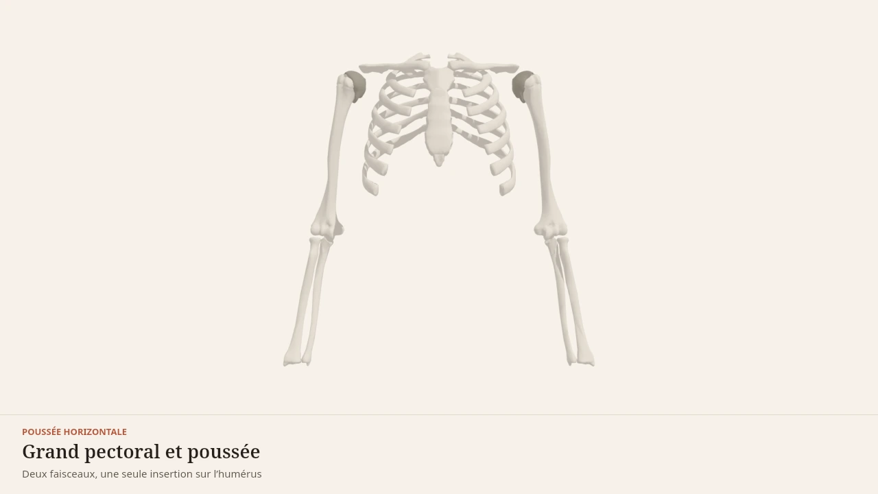
The scene highlights the muscle from the clavicle and the sternum to its attachment on the humerus. During the pushing phase, the two arms come together in front of the chest and the muscle shortens; during the descent, it lengthens while braking the movement. Without the 3D, the essential point fits in one sentence: the pectoralis major pulls the humerus forward and toward the middle of the body.
Current step
The frame of the push
Pectoralis major does not pull into thin air: it links a wide fixed base — clavicle, sternum and costal cartilages — to a single moving point on the humerus. Locating those bony attachments is already enough to understand its line of pull.
Scene description
A three-dimensional diagram of the upper trunk seen from the front, both arms held out horizontally as at the start of a bench press. The ribcage, sternum, both clavicles, both humeri and the forearms are shown in bone tones, and each shoulder joint is marked by a small dark sphere. Over this frame, pectoralis major is laid out in terracotta as two distinct heads: a thin clavicular head under the collarbone, and a much broader sternocostal head running from the sternum to the humerus, both converging on the same insertion point. The anterior deltoid caps the front of the shoulder and triceps brachii runs along the back of the arm. An animation brings the humeri in front of the chest in horizontal adduction while the belly of pectoralis major thickens. The volumes are deliberately simplified: this is a teaching diagram, not an exact anatomical reconstruction.
Visible structures
- Clavicular headThe thin upper portion of pectoralis major, arising from the medial third of the front of the clavicle. Its fibres run down to the humerus, carrying the arm forwards and upwards, and it dominates in incline pushing.
- Sternocostal headThe broad lower portion, arising from the lateral border of the sternum and the cartilages of the first six ribs. Its fibres run obliquely up to the bicipital groove of the humerus, bringing the arm forwards and downwards.
- Humeral insertionBoth heads converge into a flat tendon attached to the lateral lip of the bicipital groove of the humerus. The whole origin — clavicle, sternum and ribs — therefore pulls on this single point, which is what makes the line of pull so directional.
- Anterior deltoidThe front head of the deltoid, arising from the lateral third of the clavicle and inserting on the deltoid tuberosity. It flexes the shoulder and carries the arm forwards, working alongside pectoralis major in every push.
- Triceps brachiiThree heads joining on the olecranon of the ulna. It extends the elbow and finishes the push: without it the arm closes towards the trunk but the load never actually travels away.
Guided steps
- 1/5Pectoralis major does not pull into thin air: it links a wide fixed base — clavicle, sternum and costal cartilages — to a single moving point on the humerus. Locating those bony attachments is already enough to understand its line of pull.
- 2/5The upper portion arises from the medial third of the clavicle. Its fibres run obliquely downwards to the humerus, which gives them a flexing action: they carry the arm forwards and upwards, and dominate in incline pushing.
- 3/5The much broader lower portion arises from the sternum and the cartilages of the first six ribs. Its fibres run upwards to the humerus, bringing the arm forwards and downwards — the direction of the bench press and the push-up.
- 4/5When the arm starts out to the side and comes back in front of the chest, pectoralis major shortens and its belly thickens. This horizontal adduction is the movement it produces most efficiently, and it sits at the heart of every horizontal push.
- 5/5The anterior deltoid carries the arm forwards, and triceps brachii extends the elbow to finish the movement. None of the three is enough on its own: a horizontal push is always teamwork, which is why fatigue in a single link ends the set.
Model licence · Z-Anatomy et BodyParts3D — CC-BY-SA 4.0
The movements it takes part in
The pectoralis major is the main driver of the horizontal push: bench press, push-ups, cable press. It also contributes to the vertical push, mainly through its clavicular head, when the arm passes in front of the body before rising overhead.
It works as a brake as well. In the descent of a push-up, it lengthens while staying active to control the speed. This mode of contraction, called eccentric, is one of the stimuli studied in the literature on muscle adaptation.
Synergists
- The deltoid, through its anterior head, assists flexion and horizontal adduction.
- The triceps brachii extends the elbow while the pectoral brings the arms together.
- The serratus anterior holds the shoulder blade flat against the rib cage and gives the pectoral a stable base to pull from.
Antagonists
- The latissimus dorsi and the posterior head of the deltoid produce the opposite movement, extension and horizontal abduction.
- The middle trapezius and the rhomboids draw the shoulder blades together, the opposite of the overall action of a push.
Within the same person, these roles are not fixed: a muscle described as an antagonist can contract at the same time as the agonist to stabilise the joint.
What the 3D model simplifies
Limit
A model, not a dissection
The pectoralis major is shown as a uniform sheet with clearly separated heads. In a real body, the fibres are continuous and recruitment shifts gradually from one head to the next.
The model uses a single reference morphology. Chest width, clavicle length and the number of ribs involved in the attachment vary from person to person. No Body Lab scene therefore represents the exact anatomy of any given individual.
Sources
Key sources
- Standring S (2020). Gray's Anatomy: The Anatomical Basis of Clinical Practice, 42nd edition. Elsevier.https://shop.elsevier.com/books/grays-anatomy/standring/978-0-7020-7705-0
- Neumann DA (2016). Kinesiology of the Musculoskeletal System: Foundations for Rehabilitation, 3rd edition. Elsevier.https://shop.elsevier.com/books/kinesiology-of-the-musculoskeletal-system/neumann/978-0-323-28753-1
- Schoenfeld BJ (2010). The mechanisms of muscle hypertrophy and their application to resistance training. Journal of Strength and Conditioning Research.DOI : 10.1519/jsc.0b013e3181e840f3
Put it into practice in Shapier
Practise the dumbbell bench press
The dumbbell bench press, a horizontal push driven by the pectoralis major, is followed step by step in Shapier; Body Lab only explains the muscle's anatomy.
Practise the dumbbell bench press
Body Lab explains; Shapier lets you act and track.
Check my understanding
Which head of the pectoralis major carries the arm forward and upward?
The clavicular head
The sternocostal head
The abdominal head
Is a resistance that changes over the course of a single movement a sign of weakness?
No, it is the length–tension relationship
Yes, the muscle tires toward the end of the movement
Yes, it reveals poor execution
Can a muscle antagonist to the pectoralis major contract at the same time as it?
Yes, to stabilise the joint
No, agonist and antagonist always exclude each other
Only in trained people
Choose an answer
Read next
- DeltoidsThe deltoid caps the shoulder with three heads pointing in different directions. It raises the arm in every direction, and its shape explains why the same shoulder changes function depending on the angle of the movement.With a 3D scene
- Triceps brachiiThe triceps brachii fills the whole back of the upper arm and extends the elbow. Its long head also crosses the shoulder, which changes its length depending on the position of the arm.With a 3D scene
- Latissimus dorsiThe latissimus dorsi is the broadest muscle of the back. It links the pelvis and the spine to the humerus, and draws the arm down and backward in every pulling movement.With a 3D scene
Where to go next
- How muscle growsWhat actually makes a muscle grow? This page follows the chain of events that links a set of exercise to a thicker muscle fibre, and separates what is established from what is still debated by research.
Available offline
Offline download is available in the mobile app.
Trust and method
Author
Editorial review
Thanh Chau
Published on
August 2, 2026
Reviewed on August 2, 2026
Next review due August 2, 2027
Limits of this page
- The boundaries between the clavicular, sternocostal and abdominal heads are gradual; the 3D model separates them more sharply than dissection does.
- The extent of the costal attachments varies from person to person, and the abdominal head is sometimes very small or altogether absent.
- The page describes joint actions, not the measured contribution of each head to a given exercise.
Educational content. Body Lab does not diagnose and does not replace professional advice.
How we work