Where is the latissimus dorsi located?
The latissimus dorsi covers the lower back and links the pelvis, spine and lowest ribs to the humerus through broad attachments. It acts mainly at the shoulder, drawing the arm down, backward and toward the trunk, especially during pull-ups. When the arm is fixed, it can instead draw the trunk toward the arm.
Origins
The muscle arises from a very wide surface, largely through the thoracolumbar fascia, a fibrous sheet stretched over the lower back.
- The spinous processes of the last thoracic vertebrae and of all the lumbar vertebrae, together with the sacral region, through the thoracolumbar fascia.
- The back third of the iliac crest, the upper border of the pelvis.
- The lowest ribs, by small slips that interlock with the external oblique.
- The lower angle of the shoulder blade, inconstantly.
Insertion
All these fibres converge upward and outward, twist, and then attach by a flat tendon to the floor of the intertubercular groove of the humerus, between the attachments of the pectoralis major and the teres major.
Innervation and palpable landmarks
Innervation comes from the thoracodorsal nerve, from roots C6 to C8. The simplest landmark is the back fold of the armpit, formed by the border of the muscle: placing a hand on the side of the rib cage and drawing the elbow downward, the mass can be felt contracting.
What does the latissimus dorsi do?
The latissimus dorsi acts mainly on the shoulder, with three combined actions.
- Shoulder extension: it draws the arm backward from a forward position.
- Adduction: it brings the arm against the trunk from an abducted or raised position.
- Internal rotation: it turns the humerus inward.
It also depresses the shoulder girdle and, through its lumbar attachments, takes part in trunk movements when the arm is fixed. As an accessory, it is involved in forced expiration, during a cough for example.
Thoracolumbar fascia
A sheet of fibrous tissue stretched across the lower back, between the pelvis, the spine and the ribs. It serves as a shared attachment surface for several muscles, including the latissimus dorsi, and transmits forces from one side of the body to the other.
lumbodorsal aponeurosis
Seeing the latissimus dorsi in motion
The latissimus dorsi during a pull
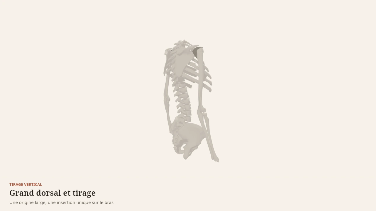
The scene shows the muscle from its lumbar attachments to the humerus, during a vertical pull. As the elbow travels down, the fibres shorten in a fan and the shoulder drops. Without the 3D, remember this: the latissimus dorsi pulls the arm down and backward, taking its support from the pelvis and the spine.
Current step
Three parts to connect
A pull connects the hand to the pelvis through three moving parts: the humerus, the shoulder blade gliding over the ribcage, and the spine acting as the fixed point. Latissimus dorsi is the only muscle to span almost that whole path, from pelvis to arm.
Scene description
A three-dimensional diagram of the trunk seen from behind, arms raised diagonally overhead as at the start of a vertical pull. The ribcage, the spine, the pelvis, both shoulder blades, both humeri and the forearms are shown in bone tones, with a small dark sphere marking each shoulder joint. In terracotta, latissimus dorsi forms a wide fan on each side, spreading from the lower back and iliac crest and narrowing as it rises to the top of the humerus. The trapezius is split into three bands — upper towards the shoulder, middle towards the spine of the scapula, lower rising from the mid back — and biceps brachii runs along the front of the arm. A two-and-a-half-second animation brings the humeri back alongside the trunk in adduction while the shoulder blades descend and latissimus dorsi shortens. The volumes are stylised: this is a teaching diagram, not an exact anatomical reconstruction.
Visible structures
- Latissimus dorsiA broad fan arising from the spinous processes of the lower thoracic and lumbar vertebrae, the thoracolumbar fascia, the iliac crest and the last three or four ribs. All its fibres converge on the bicipital groove of the humerus: it pulls the arm down, back and towards the trunk.
- Shoulder bladeA flat bone gliding over the back of the ribcage, with no direct joint to the spine. In a pull it depresses and rotates downwards: without that motion the humerus jams against the acromion and the range gets paid for elsewhere.
- TrapeziusIts three portions pull the shoulder blade in three directions: the upper elevates it, the middle draws it towards the spine, the lower depresses it. In a vertical pull, the lower portion is the direct partner of latissimus dorsi.
- Biceps brachiiTwo heads arising from the scapula and inserting on the radial tuberosity. It flexes the elbow and supinates the forearm: in a pull it shortens the distance between hand and shoulder while latissimus dorsi moves the arm.
- Insertion on the humerusThe tendon of latissimus dorsi attaches in the floor of the bicipital groove, at the very top of the humerus. A wide origin across the whole lower back and a narrow insertion high on the arm: that convergence is what gives the muscle its fan shape.
Guided steps
- 1/5A pull connects the hand to the pelvis through three moving parts: the humerus, the shoulder blade gliding over the ribcage, and the spine acting as the fixed point. Latissimus dorsi is the only muscle to span almost that whole path, from pelvis to arm.
- 2/5Latissimus dorsi anchors across almost the whole lower back: the lower thoracic and lumbar spinous processes, the thoracolumbar fascia, the iliac crest and the lowest ribs. That origin surface is what gives it its strength, and why a pull is felt all the way down to the pelvis.
- 3/5All those fibres converge into a flat tendon attached in the floor of the bicipital groove of the humerus. Wide below, narrow above: the pull concentrates on a single point of the arm, which makes it strongly directional — downwards and backwards.
- 4/5The humeri leave the overhead position and adduct back alongside the trunk while the shoulder blades depress and rotate downwards. Latissimus dorsi shortens, the lower trapezius holds the blade down and the elbow bends: arm motion and shoulder-blade motion cannot be separated.
- 5/5The lower trapezius depresses the shoulder blade, the middle trapezius draws it towards the spine, and biceps brachii flexes the elbow to bring the hand closer to the shoulder. A pull is never an isolated back exercise: it is coordination between arm, shoulder blade and spine.
Model licence · Z-Anatomy et BodyParts3D — CC-BY-SA 4.0
Which movements involve the latissimus dorsi?
The latissimus dorsi is the main driver of the vertical pull: pull-up, lat pulldown. It is also strongly engaged in the horizontal pull, where shoulder extension dominates over adduction.
It is involved more indirectly in the hip hinge: when the arms hold a load, the muscle keeps the bar close to the body and transmits part of the forces toward the thoracolumbar fascia.
Synergists
- The teres major, often described as its assistant, produces the same shoulder actions over a shorter range.
- The posterior head of the deltoid assists shoulder extension.
- The biceps brachii and the brachialis flex the elbow while the latissimus draws the arm in.
- The lower trapezius depresses and stabilises the shoulder blade.
Antagonists
- The middle head of the deltoid and the supraspinatus take the arm away from the body, the opposite of adduction.
- The pectoralis major, through its clavicular head, carries the arm forward.
- The external rotators of the shoulder oppose its internal rotation.
What the 3D model simplifies
Limit
A fibrous sheet reduced to a surface
The thoracolumbar fascia is displayed as a single membrane. In reality it is made of several stacked layers that receive fibres from different muscles and that take part in transmitting forces between the trunk and the limbs.
The model also does not show the real overlap with the trapezius and the erector spinae, which occupy the same anatomical space at different depths. The extent of the costal attachments and the presence of the scapular attachment vary from person to person.
Sources
Key sources
- Standring S (2020). Gray's Anatomy: The Anatomical Basis of Clinical Practice, 42nd edition. Elsevier.https://shop.elsevier.com/books/grays-anatomy/standring/978-0-7020-7705-0
- Neumann DA (2016). Kinesiology of the Musculoskeletal System: Foundations for Rehabilitation, 3rd edition. Elsevier.https://shop.elsevier.com/books/kinesiology-of-the-musculoskeletal-system/neumann/978-0-323-28753-1
Put it into practice in Shapier
See the pull-up in Shapier
Open in Shapier the pull-up exercise page, the vertical pull the latissimus dorsi mainly drives, to put it into practice.
See the pull-up in Shapier
Body Lab explains; Shapier lets you act and track.
Check my understanding
During a pull-up, with the arm fixed, what does the latissimus dorsi do?
It draws the trunk toward the arm
It cannot contract, for lack of a moving segment
It flexes the elbow to lift the body
In a horizontal pull, which action of the latissimus dorsi dominates?
Shoulder extension
Adduction, as in every pull
Internal rotation
What does the 3D model simplify about the thoracolumbar fascia?
It displays it as a single membrane, whereas it is made of several stacked layers
It omits it from the scene entirely
It shows it attached only to the pelvis
Choose an answer
Read next
- TrapeziusThe trapezius links the skull and the spine to the shoulder girdle. Its three portions orient the shoulder blade, a condition without which the arm cannot rise fully.With a 3D scene
- Biceps brachiiThe biceps brachii sits on the front of the upper arm and crosses both the shoulder and the elbow. It flexes the elbow, but its most specific function is supination of the forearm.With a 3D scene
- Erector spinaeThe erector spinae form three muscular columns running along the spine. They straighten it, control its forward bending and hold the trunk during hip hinge movements.With a 3D scene
Where to go next
- How muscle growsWhat actually makes a muscle grow? This page follows the chain of events that links a set of exercise to a thicker muscle fibre, and separates what is established from what is still debated by research.
Available offline
Offline download is available in the mobile app.
Trust and method
Author
Editorial review
Thanh Chau
Published on
August 2, 2026
Reviewed on August 2, 2026
Next review due August 2, 2027
Limits of this page
- The thoracolumbar fascia, through which the muscle attaches to the pelvis and the spine, is drawn as a single surface whereas it is a fibrous sheet made of several layers.
- The attachment on the lower angle of the shoulder blade is inconstant and is not drawn separately.
- The page describes joint actions, not how the work is shared between the latissimus dorsi and the teres major in a given pull.
Educational content. Body Lab does not diagnose and does not replace professional advice.
How we work