Where the erector spinae are located
The erector spinae are not a single muscle but a set of bundles arranged in three vertical columns, on each side of the spine. They fill the groove between the spinous processes, in the middle of the back, and the ribs. This is the relief felt on either side of the midline, from the lower back up to the neck.
Common origin
At the bottom, the three columns start from a thick common tendinous mass, attached to:
- the back surface of the sacrum and the sacroiliac ligaments;
- the inner part of the iliac crest;
- the spinous processes of the lumbar vertebrae and of the last thoracic vertebrae.
The three columns
From the outside inward, three sets can be distinguished, relaying one another like a staircase along the back.
- The iliocostalis, the most lateral, reaches the ribs.
- The longissimus, intermediate and the longest, reaches the transverse processes of the vertebrae and, for its highest bundle, the mastoid process of the skull.
- The spinalis, the most medial, links spinous processes to one another.
Innervation and palpable landmarks
These muscles are supplied by the posterior branches of the spinal nerves, at every level of the spine. The simplest landmark is the double column felt on either side of the spinous processes, particularly clear in the lumbar region as the trunk straightens.
What the erector spinae do
- Spinal extension: contracting on both sides, they straighten the spine and draw the trunk backward.
- Control of forward bending: as the trunk tips forward, they lengthen while staying active and slow the descent.
- Side bending: contracting on one side only, they tilt the trunk to that side.
- Contribution to rotation: they accompany rotation of the trunk together with the obliques and the deep muscles.
Eccentric contraction
A contraction during which the muscle produces tension while lengthening. It is the way the erectors work when the trunk lowers forward under control. The three modes are compared on the [contraction regimes](/en/body/contraction-regimes) page.
braking work
Certainty level · Probable
Trunk stability depends on the coordinated action of several muscles rather than on any single one.
Spine biomechanics research describes the spinal column as a structure whose stiffness increases with co-contraction of the surrounding muscles, erectors included. This model is consistent with laboratory measurements, but moving from those measurements to training recommendations remains debated: the protocols differ, the populations studied are heterogeneous, and the reported effects cannot be transferred as they stand to a given person.
Neumann DA (2016) · McGill SM (2010)
Seeing the erectors in motion
The erector spinae during a hip hinge
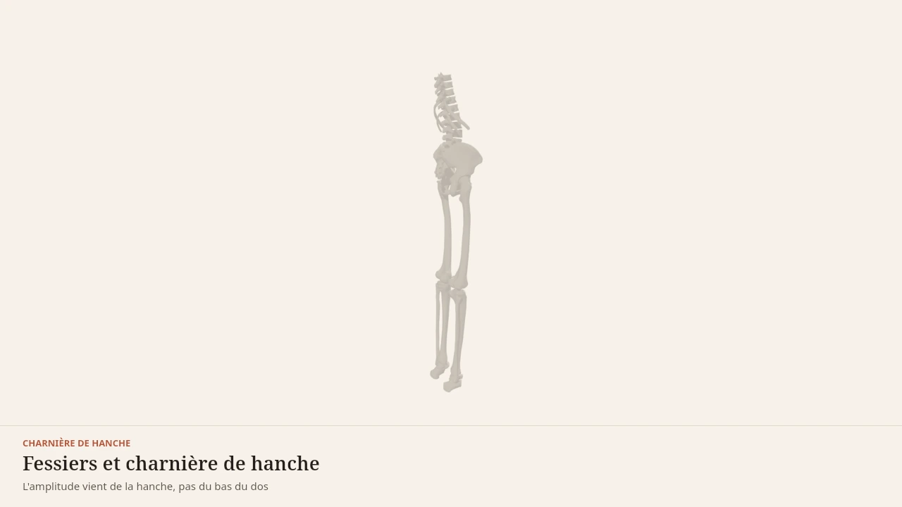
The scene shows the back while the pelvis travels backward and the trunk tips forward. The muscular columns stay active without the spine rounding: the movement comes from the hip. Without the 3D, the idea is that these muscles serve here to hold the position of the back rather than to produce the movement.
Current step
A single pivot
The hip hinge plays out between three parts: the pelvis, the lumbar spine resting on it, and the femur articulating beneath. The only useful pivot is the hip, where the femoral head turns in the acetabulum. The lumbar spine is not built to supply the range.
Scene description
A three-dimensional diagram of the lower half of the body seen from behind, standing with the legs straight. The pelvis, both femurs, both tibias and a simplified lumbar spine — five vertebral bodies with their spinous processes — are shown in bone tones, while the feet and a reference trunk stay in a neutral paper shade. In terracotta, gluteus maximus caps the back of the pelvis, the hamstrings run down behind each thigh in two bundles — biceps femoris outside, semitendinosus inside — and two columns of erector spinae flank the spine up to the mid back. A three-second animation tips the pelvis forwards around the hips: the buttocks travel back, the trunk leans over, the knees bend only slightly and the hamstrings lengthen, before the movement returns to standing. The volumes are stylised: this is a teaching diagram, not an exact anatomical reconstruction.
Visible structures
- Gluteus maximusThe bulkiest muscle in the body, arising from the back of the iliac wing, the sacrum and the sacrotuberous ligament, and inserting on the gluteal tuberosity of the femur and the iliotibial tract. It extends the hip powerfully, above all at the end of the lockout.
- HamstringsBiceps femoris on the outside, semitendinosus and semimembranosus on the inside, all arising from the ischial tuberosity and inserting on the fibula and tibia. They extend the hip and flex the knee: in the hinge they lengthen while braking the descent.
- Erector spinaeMuscular columns — iliocostalis, longissimus and spinalis — running from the sacrum and iliac crest to the ribs and vertebrae. In the hinge they do not produce the movement: they keep the lumbar spine from rounding while the hip flexes.
- Lumbar spineFive vertebrae stacked between the sacrum and the ribs, shown here as their bodies and spinous processes. A sound hinge rotates the pelvis on the hips while these five levels stay almost still relative to one another.
- The hip jointThe femoral head turns inside the acetabulum of the pelvis: this is the only pivot of the movement. The whole point of the hinge is to rotate the pelvis around this axis rather than to bend the back.
Guided steps
- 1/5The hip hinge plays out between three parts: the pelvis, the lumbar spine resting on it, and the femur articulating beneath. The only useful pivot is the hip, where the femoral head turns in the acetabulum. The lumbar spine is not built to supply the range.
- 2/5It arises behind the iliac wing, from the sacrum and the sacrotuberous ligament, and reaches the femur and the iliotibial tract. Its bulk and lever arm make it the main hip extensor, especially through the last part of standing back up.
- 3/5All three set off from the ischial tuberosity and run down behind the thigh to the tibia and fibula. Because they cross both hip and knee, they lengthen further the more the pelvis tips forward with the knees nearly straight.
- 4/5The pelvis rotates forwards around the hips, the buttocks travel backwards, and the knees bend only slightly. The hamstrings lengthen while braking the descent, then the glutes bring the pelvis back upright. The trunk follows the pelvis without the back changing shape.
- 5/5Throughout the tip, the erector spinae work without shortening: they stop the spine from rounding. That is how a hinge is judged — the range comes from the hip, the spine keeps its shape, and force travels from the legs to the trunk without leaking.
Model licence · Z-Anatomy et BodyParts3D — CC-BY-SA 4.0
The movements they take part in
The erector spinae are central to the hip hinge: they hold the trunk while the glutes and the hamstrings produce hip extension. They work the same way in the squat, where they keep the trunk from tipping forward under the load.
They are also involved in movements where the load is held away from the body, such as the horizontal pull performed with the trunk bent forward.
Synergists
- The multifidus, a deep muscle, controls the orientation of each vertebra.
- The quadratus lumborum takes part in side bending and stabilises the twelfth rib.
- The glutes and the hamstrings extend the hip while the erectors hold the trunk.
- The latissimus dorsi shares the thoracolumbar fascia with them.
Antagonists
- The abdominal wall flexes the trunk, the opposite action to extension.
- The psoas and the iliacus, hip flexors, change the orientation of the pelvis and therefore the position of the lumbar spine.
What the 3D model simplifies
Limit
Columns more continuous than they appear
The model clearly separates the iliocostalis, the longissimus and the spinalis. In dissection, these bundles overlap, exchange fibres and continue into one another: the separation is a reading aid, not an observable reality.
The scene does not show the deep muscles lying beneath these columns, nor the thoracolumbar fascia at its real thickness. The number of lumbar vertebrae and the shape of the spinal curves vary from person to person, and the model represents a reference morphology.
Sources
Key sources
- Standring S (2020). Gray's Anatomy: The Anatomical Basis of Clinical Practice, 42nd edition. Elsevier.https://shop.elsevier.com/books/grays-anatomy/standring/978-0-7020-7705-0
- Neumann DA (2016). Kinesiology of the Musculoskeletal System: Foundations for Rehabilitation, 3rd edition. Elsevier.https://shop.elsevier.com/books/kinesiology-of-the-musculoskeletal-system/neumann/978-0-323-28753-1
- McGill SM (2010). Core training: evidence translating to better performance and injury prevention. Strength and Conditioning Journal.DOI : 10.1519/ssc.0b013e3181df4521
Put it into practice in Shapier
Practice the Romanian deadlift
Open in Shapier the Romanian deadlift guide, the hip hinge where the erectors hold the trunk.
Practice the Romanian deadlift
Body Lab explains; Shapier lets you act and track.
Check my understanding
During a hip hinge, what is the main role of the erector spinae?
Holding the trunk while the glutes and hamstrings produce hip extension
Producing hip extension by driving the pelvis upright
Gradually relaxing as the trunk tips forward
What do the erector spinae do as the trunk tips forward?
They lengthen while staying active and slow the descent
They switch off since the spine is flexing
They shorten to pull the trunk backward right away
What should be remembered about the split into three columns, iliocostalis, longissimus and spinalis?
It is a reading aid: in dissection, the bundles overlap and continue into one another
They are three independent muscles with sharp boundaries
Only the longissimus really exists, the other two are diagrams
Choose an answer
Read next
- Abdominal wallThe abdominal wall brings together four stacked muscles whose fibres cross. It flexes and rotates the trunk, but its most constant role is to resist movement and to manage internal pressure.With a 3D scene
- GlutesThe three gluteal muscles cover the back and the side of the pelvis. The gluteus maximus extends the hip, while the gluteus medius and minimus steady the pelvis as soon as the body is supported on one leg.With a 3D scene
- HamstringsThe hamstrings occupy the back of the thigh. They extend the hip and flex the knee, and their length depends on the combined position of these two joints.With a 3D scene
Where to go next
- How muscle growsWhat actually makes a muscle grow? This page follows the chain of events that links a set of exercise to a thicker muscle fibre, and separates what is established from what is still debated by research.
Available offline
Offline download is available in the mobile app.
Trust and method
Author
Editorial review
Thanh Chau
Published on
August 2, 2026
Reviewed on August 2, 2026
Next review due August 2, 2027
Limits of this page
- The 3D model shows three distinct columns, whereas the bundles overlap widely and continue into one another with no sharp boundary.
- The deep muscles of the back, in particular the multifidus and the rotatores, are not shown even though they take part in segmental control of the spine.
- This page describes anatomy and function; it covers neither low back pain nor what to do when a symptom appears.
Educational content. Body Lab does not diagnose and does not replace professional advice.
How we work