What is the squat used for?
The squat lets the body lower and rise by coordinating the hips, knees and ankles while the feet stay on the ground. This pattern predates the gym: sitting down, standing from a low seat, and picking up an object while keeping the torso close to vertical all rely on the same movement.
Its mechanical signature is that it is a movement in a closed kinetic chain. The foot never leaves the ground: the three joints of the lower limb are therefore obliged to coordinate, and none of them can move on its own without the other two adjusting. That constraint is what makes the squat a whole-body movement rather than a thigh exercise (neumann2016).
Eccentric phase
Phase during which a muscle produces force while lengthening under the load. In the squat it corresponds to the descent: the knee and hip extensors slow the movement down instead of producing it. The three [contraction regimes](/en/body/contraction-regimes) are described together elsewhere.
eccentric contraction · lowering phase
What are the phases of a squat?
Breaking the squat into phases serves to distinguish moments in which the same muscles do not play the same role. The sequence below describes a squat performed at a controlled speed.
The squat, phase by phase
- 1Bracingbefore the descentThe feet take support, the ribcage and the pelvis line up, and the trunk muscles contract together to stiffen the spine before the load starts moving.
- 2Eccentric phasedescentHips, knees and ankles flex together. The knee and hip extensors work while lengthening: they slow the descent rather than produce it.
- 3Transitionbottom positionThe movement reverses. This is the moment when the lever arms are longest and when the demand on the hip and knee extensors is highest.
- 4Concentric phaseascentThe same muscles shorten and extend the three joints. Hips and knees have to extend at the same rate, failing which the torso tips forward.
- 5Lockoutend of the ascentThe hips and knees complete their extension, the pelvis returns underneath the ribcage, and breathing resumes before the next repetition.
Joints and planes of movement
The squat takes place mainly in the sagittal plane, the one that divides the body into a left half and a right half: flexion and extension are visible there from the side.
- Hip: a ball-and-socket joint between the femoral head and the acetabulum. It flexes on the
way down, extends on the way up, and simultaneously controls adduction and rotation in the two other planes (standring2020).
- Knee: a tibiofemoral joint working in flexion and extension, paired with a patellofemoral
joint in which the kneecap slides in the groove of the femur. Patellofemoral compression increases with the degree of flexion (escamilla2001).
- Ankle: dorsiflexion at the talocrural joint allows the shin to travel forward over the
foot. Its range directly conditions how far the torso leans.
- Lumbar spine: it does not produce the movement, it is held in a relatively stable position
while the hips and knees work (mcgill2010).
Which muscles work during a squat?
Muscles are not divided into “useful” and “useless” ones but into roles: producing the movement, contributing to it, or holding a segment in place.
Muscle roles in the squat
Role
Main structures
What they do
Prime movers
Quadriceps, gluteus maximus
Extend the knee and the hip during the ascent, slow their flexion during the descent.
Synergists
Hamstrings, adductor magnus, calves
Complete hip extension and stabilise the knee and the ankle during the drive.
Stabilisers
Erector spinae, abdominal wall, gluteus medius
Hold the trunk and the pelvis while the lower limbs move.
The hamstrings occupy a special place: they cross both the hip and the knee. They flex the knee and extend the hip at once, so that their overall length varies little during the squat. Their work there is therefore largely stabilising, very different from what they supply in a hip hinge (escamilla2001).
Certainty level · Established
Squat depth changes how loading is distributed between the knee and the hip; it does not in itself constitute a factor of joint injury.
Biomechanical work on the squat shows that tibiofemoral and patellofemoral compressive forces increase as the knee flexes, while the shear stress borne by the cruciate ligaments stays low across the whole range. These measurements describe internal forces, not injuries: they come from motion analyses and models, in a limited number of trained participants. The step from “higher loading” to “risk to the joint” is not demonstrated by this work.
Escamilla RF (2001) · Schoenfeld BJ (2010)
What morphology changes
Two people do not squat in the same way, and that comes first of all from anatomy. The ratio between femur length and trunk length determines how far the torso has to lean to keep the whole system balanced over the foot: the longer the femur, the more the torso has to lean, which increases the demand on the hip extensors (schoenfeld2010squat).
Acetabular orientation and the angle of the femoral neck also vary between individuals (standring2020): a stance width that is comfortable for one person is not necessarily so for another. Ankle dorsiflexion range plays the same part lower down: when it is limited, the knee travels forward less and the torso leans more.
Technical variations follow the same logic. Placing the load in front of the body rather than behind the neck forces a more upright torso and shifts the demand towards the knee extensors; the reverse loads the hip extensors more (schoenfeld2010squat).
Common misunderstandings
Caution
Three misconceptions about the squat
“The knee must never travel past the toes.” Limiting forward travel of the knee does reduce the moment at the knee, but it transfers the demand to the hip and the lower back. This is a redistribution of loading, not its removal.
“Deep squats wreck the knees.” Biomechanical analyses measure internal forces that vary with the angle of flexion; they do not measure tissue damage. The depth that can be reached depends above all on the available joint ranges.
“You have to keep your back straight.” What the literature describes is not a vertical spine but a spine held in a stable position during effort. A torso leaning forward is not the same thing as a flexed spine.
A fourth confusion is worth clearing up: the squat is not a quadriceps exercise in which the glutes take part incidentally. The respective contribution of the hip and knee extensors shifts with depth and with how far the torso leans (schoenfeld2010squat).
Quadriceps and the squat
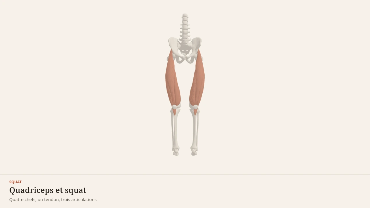
The model shows the lower limb during the descent and the ascent, with the knee and hip extensors highlighted in turn. The text above stays complete without the scene: the scene illustrates the coordination between hip, knee and ankle, and represents nobody's individual morphology.
Current step
Three segments, three joints
The squat is not a thigh movement: it is a sequence. Pelvis, femur and tibia form three segments linked by the hip, the knee and the ankle, with the patella sitting in front of the knee. The whole mechanics of the movement lives in those pivots.
Scene description
A three-dimensional diagram of the lower half of the body, standing, seen from the front: pelvis, femurs, tibias and patellae are shown in bone tones, with the feet and a reference trunk in a neutral paper shade. On each thigh the four heads of the quadriceps are laid over in terracotta and stay distinct: rectus femoris in the middle, vastus lateralis on the outside, vastus medialis on the inside, and the deeper vastus intermedius against the femur. Behind, the glutes cap the pelvis and the calves fill the upper shank. A three-second animation flexes the ankle, knee and hip together: the knee travels forward over the foot, the hip moves back and down, the trunk leans, and then the movement reverses. The volumes are stylised: this is a teaching diagram, not an exact anatomical reconstruction.
Visible structures
- Rectus femorisThe only head of the quadriceps to cross two joints: it arises from the anterior inferior iliac spine of the pelvis and runs down the middle of the thigh into the quadriceps tendon. It extends the knee and flexes the hip.
- Vastus lateralisThe largest of the four heads, arising from the greater trochanter and the linea aspera on the outer side of the femur. It acts on the knee alone, extending it, and gives the thigh its outer contour.
- Vastus medialisThe inner head, arising from the linea aspera of the femur, whose lowest fibres reach the patella almost horizontally. It extends the knee and helps keep the patella tracking in line.
- Vastus intermediusThe deep head, pressed against the front of the femur and covered by the other three. Invisible from the outside, it still produces a substantial share of knee extension.
- PatellaA sesamoid bone embedded in the quadriceps tendon, which continues as the patellar tendon onto the tibial tuberosity. By holding the tendon away from the knee axis, it increases the lever arm of the quadriceps.
- GlutesArising from the iliac wing and the sacrum, inserting on the femur and the iliotibial tract. In the squat they extend the hip: the more the trunk leans forward, the larger their share of the work compared with the quadriceps.
- CalvesThe gastrocnemius and soleus, joining on the calcaneus through the Achilles tendon. In the squat they work mostly as brakes, controlling ankle dorsiflexion and therefore how far the knee travels forward.
Guided steps
- 1/5The squat is not a thigh movement: it is a sequence. Pelvis, femur and tibia form three segments linked by the hip, the knee and the ankle, with the patella sitting in front of the knee. The whole mechanics of the movement lives in those pivots.
- 2/5Rectus femoris occupies the middle of the thigh, vastus lateralis its outer border, vastus medialis its inner border, and vastus intermedius hides beneath them against the femur. All four converge on a single tendon: they pull together, but only rectus femoris also crosses the hip.
- 3/5The quadriceps tendon wraps around the patella and continues to the tibial tuberosity. By holding the tendon away from the axis of flexion, the patella lengthens the lever arm — that is what makes knee extension effective in the bottom position.
- 4/5Ankle, knee and hip flex together. The ankle lets the knee travel forward over the foot, the femur tips backwards, and the trunk leans to keep balance over the midfoot. The quadriceps first brakes the descent, then drives the way back up.
- 5/5The glutes extend the hip and take on more work the further the trunk leans; the calves restrain the ankle and steady the base. The quadriceps is the most heavily loaded engine in the squat, but it does not decide the depth reached on its own.
Model licence · Z-Anatomy et BodyParts3D — CC-BY-SA 4.0
The squat forms a pair with the hip hinge: the first shares the work between hip and knee, the second concentrates it at the hip.
The pages devoted to the quadriceps, the glutes, the hamstrings and the erector spinae detail the attachments and the functions summarised here.
Key sources
- Neumann DA (2016). Kinesiology of the Musculoskeletal System: Foundations for Rehabilitation, 3rd edition. Elsevier.https://shop.elsevier.com/books/kinesiology-of-the-musculoskeletal-system/neumann/978-0-323-28753-1
- Standring S (2020). Gray's Anatomy: The Anatomical Basis of Clinical Practice, 42nd edition. Elsevier.https://shop.elsevier.com/books/grays-anatomy/standring/978-0-7020-7705-0
- Escamilla RF (2001). Knee biomechanics of the dynamic squat exercise. Medicine and Science in Sports and Exercise.DOI : 10.1097/00005768-200101000-00020
- Schoenfeld BJ (2010). Squatting kinematics and kinetics and their application to exercise performance. Journal of Strength and Conditioning Research.DOI : 10.1519/jsc.0b013e3181bac2d7
- McGill SM (2010). Core training: evidence translating to better performance and injury prevention. Strength and Conditioning Journal.DOI : 10.1519/ssc.0b013e3181df4521
Put it into practice in Shapier
Open the squat exercise page
Find in Shapier the squat exercise page, to place the movement within a session.
Open the squat exercise page
Body Lab explains; Shapier lets you act and track.
Check my understanding
What happens when the knee is prevented from travelling past the toes?
The loading is redistributed to the hip and the lower back
The knee is completely unloaded
The quadriceps work harder
What do biomechanical analyses of the deep squat actually measure?
Internal forces that vary with the angle of flexion, with no measure of tissue damage
Knee injuries caused by deep flexion
Cartilage wear proportional to squat depth
What is the hamstrings' role in the squat?
A largely stabilising role, because their overall length varies little
Prime movers of the ascent
No role, since they do not produce the flexion
Choose an answer
Read next
- Hip hingeThe hip hinge concentrates the movement at the hip: the pelvis tilts over the femoral heads, the torso leans forward, and the knees stay only slightly bent. This page describes its phases, its joints, its muscles and the confusions that surround it.With a 3D scene
- QuadricepsThe quadriceps brings together four muscles on the front of the thigh that share a common tendon. It extends the knee, and one of its heads also crosses the hip.With a 3D scene
- GlutesThe three gluteal muscles cover the back and the side of the pelvis. The gluteus maximus extends the hip, while the gluteus medius and minimus steady the pelvis as soon as the body is supported on one leg.With a 3D scene
- How muscle growsWhat actually makes a muscle grow? This page follows the chain of events that links a set of exercise to a thicker muscle fibre, and separates what is established from what is still debated by research.With a 3D scene
Available offline
Offline download is available in the mobile app.
Trust and method
Author
Editorial review
Thanh Chau
Published on
August 2, 2026
Reviewed on August 2, 2026
Next review due August 2, 2027
Limits of this page
- The muscle descriptions correspond to a reference anatomical layout; muscle shape, the orientation of the femoral neck and that of the acetabulum vary from one person to another and are not represented here.
- The phases described correspond to a squat performed with a vertical external load at a controlled speed; a jump squat, a very fast squat or a markedly asymmetrical one distributes the loading differently.
- This page explains a movement, it assesses no individual execution: persistent pain, a joint that locks, or discomfort that returns at every session are matters for a healthcare professional.
Educational content. Body Lab does not diagnose and does not replace professional advice.
How we work