What the lunge is for
The squat shares the load across two symmetrical supports. The lunge puts one foot in front, one behind, and lowers the body between them. The difference is not only one of range: it changes the nature of the problem the body has to solve.
In a squat, the body has to produce force. In a lunge, it has to produce force and hold a sideways balance that nothing guarantees. That second task calls on muscles which, in the squat, stay in the background.
Unilateral stance
A configuration in which the two feet are neither side by side nor loaded the same way. The base of support becomes narrow from side to side, and controlling the pelvis in that plane becomes a constraint in its own right.
staggered stance
The phases of the movement
The lunge, phase by phase
- 1Starting position0 sStaggered stance, weight split between the ball of the back foot and the whole front foot.
- 2Descenteccentric phaseFront hip and knee flex, the back knee travels towards the floor; the muscles brake while lengthening.
- 3Bottom positiontransitionThe trunk is close to vertical, the pelvis low; sideways control is most loaded here.
- 4Riseconcentric phaseExtension of the front hip and knee, pushing into the floor; the body rises without the pelvis giving way sideways.
Joints and planes of movement
The lunge moves the same joints as the squat — hip, knee, ankle — but adds a constraint in a plane the squat barely loads.
- Sagittal plane: flexion and extension of hip and knee, as in the squat. That is the visible movement.
- Frontal plane: the pelvis tends to drop towards the back leg, which carries less load. Controlling that drop is what sets the lunge apart.
- Transverse plane: the femur of the front leg tends to turn inwards; the hip rotators resist that.
Who does the work
Muscle roles in the lunge
Role
Front leg
Back leg
Prime mover
Quadriceps and gluteus maximus
Modest share of extension
Frontal stabilisation
Gluteus medius and adductors
Adductors, holding back
Pelvic control
Erector spinae and abdominal wall
Iliopsoas, stretched by the open hip
Ankle control
Tibialis anterior and calf
Calf, in a lengthened position
Two muscles described elsewhere take on an importance here that they lack in the squat. The hip adductors control sideways movement of the pelvis, a task that becomes central as soon as the base narrows. The iliopsoas of the back leg ends up in a lengthened position, with the hip open: it is that position, not any deliberate stretch, that explains the sensation felt at the front of the rear thigh.
Certainty level · Established
Knee flexion under load distributes stress between the patellofemoral joint and the structures of the knee, according to the angle reached and the trunk position.
Reference kinesiology and work on squat biomechanics describe that distribution. Transferring it to the lunge is reasonable for the joint itself, less so for the split between the two legs, which that work does not study.
Neumann DA (2016) · Escamilla RF (2001)
Certainty level · Probable
Trunk control contributes to force transmission in movements on a reduced base of support.
The review on core training describes the role of the muscles framing the spine in transmitting force between lower and upper limbs. The size of their contribution in a lunge specifically is not established separately, and depends on load, speed and range.
McGill SM (2010)
What body proportions change
The relative length of femur and tibia markedly changes what is reachable. A long femur requires a longer step to reach the same depth, which tips the trunk further forward and shifts the demand towards the hip. The available ankle dorsiflexion range sets how far the front knee can travel.
These variations are anatomical, not technical. A position achieved by someone whose segments are proportioned differently is no benchmark.
Common misreadings
Caution
Three beliefs about the lunge
That it is a squat on one leg. The difference lies in the frontal plane: the lunge demands sideways control that the squat does not. The muscles involved are not merely fewer, they are not the same.
That the knee should never pass the toes. How far the knee travels depends on the available ankle range and on segment lengths. The load shifts between joints; it does not disappear if you prevent that travel.
That the sensation at the front of the rear thigh is a stretch to chase. It comes from the open position of the hip. This page describes what happens; it does not say what should be done.
Seeing the movement
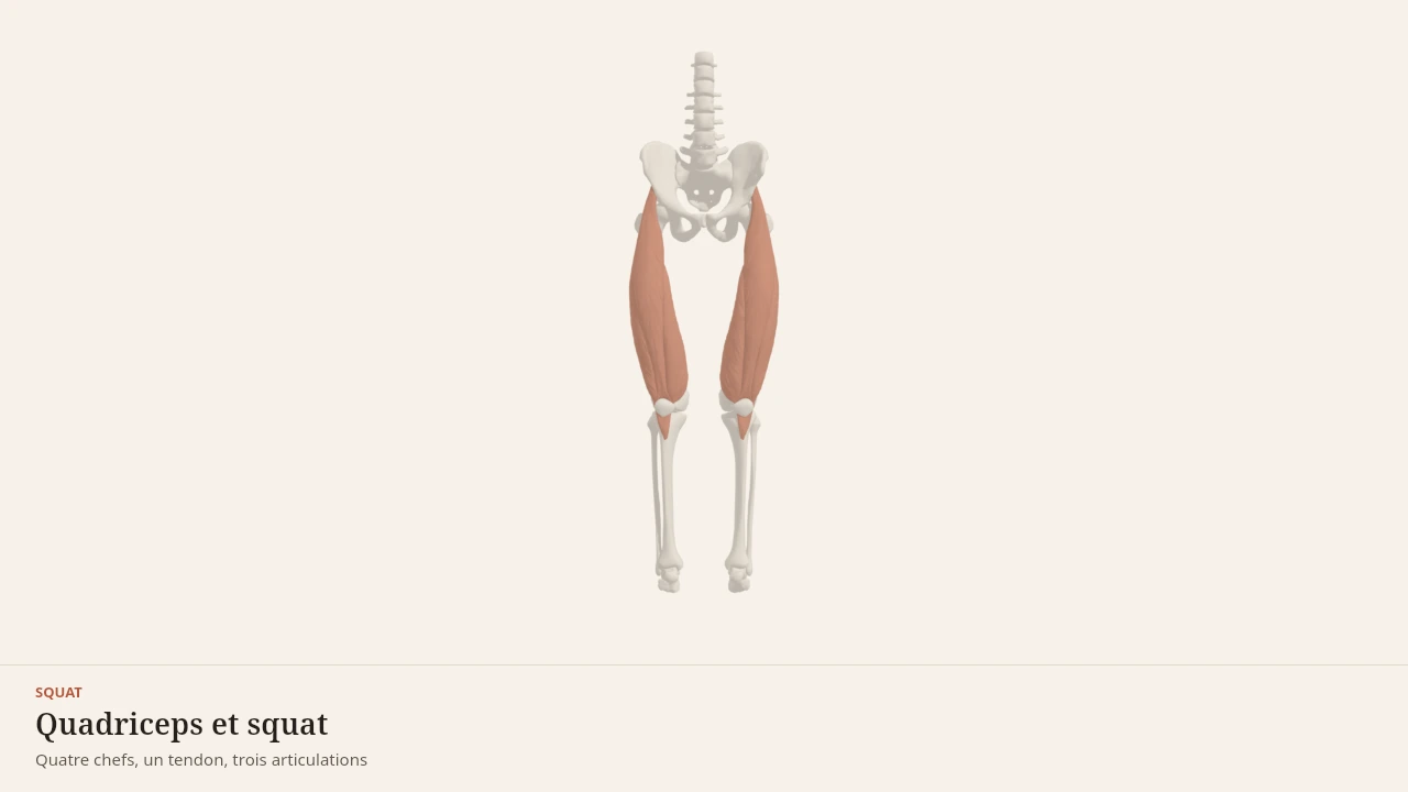
Quadriceps and squat
The scene shows the quadriceps working during a loaded knee bend. The lunge calls on the same prime movers in the front leg, adding a sideways control the scene does not depict. Without the 3D, the essentials fit in one sentence: knee and hip close on the way down and open on the way up, on a narrow base.
Current step
Three segments, three joints
The squat is not a thigh movement: it is a sequence. Pelvis, femur and tibia form three segments linked by the hip, the knee and the ankle, with the patella sitting in front of the knee. The whole mechanics of the movement lives in those pivots.
Scene description
A three-dimensional diagram of the lower half of the body, standing, seen from the front: pelvis, femurs, tibias and patellae are shown in bone tones, with the feet and a reference trunk in a neutral paper shade. On each thigh the four heads of the quadriceps are laid over in terracotta and stay distinct: rectus femoris in the middle, vastus lateralis on the outside, vastus medialis on the inside, and the deeper vastus intermedius against the femur. Behind, the glutes cap the pelvis and the calves fill the upper shank. A three-second animation flexes the ankle, knee and hip together: the knee travels forward over the foot, the hip moves back and down, the trunk leans, and then the movement reverses. The volumes are stylised: this is a teaching diagram, not an exact anatomical reconstruction.
Visible structures
- Rectus femorisThe only head of the quadriceps to cross two joints: it arises from the anterior inferior iliac spine of the pelvis and runs down the middle of the thigh into the quadriceps tendon. It extends the knee and flexes the hip.
- Vastus lateralisThe largest of the four heads, arising from the greater trochanter and the linea aspera on the outer side of the femur. It acts on the knee alone, extending it, and gives the thigh its outer contour.
- Vastus medialisThe inner head, arising from the linea aspera of the femur, whose lowest fibres reach the patella almost horizontally. It extends the knee and helps keep the patella tracking in line.
- Vastus intermediusThe deep head, pressed against the front of the femur and covered by the other three. Invisible from the outside, it still produces a substantial share of knee extension.
- PatellaA sesamoid bone embedded in the quadriceps tendon, which continues as the patellar tendon onto the tibial tuberosity. By holding the tendon away from the knee axis, it increases the lever arm of the quadriceps.
- GlutesArising from the iliac wing and the sacrum, inserting on the femur and the iliotibial tract. In the squat they extend the hip: the more the trunk leans forward, the larger their share of the work compared with the quadriceps.
- CalvesThe gastrocnemius and soleus, joining on the calcaneus through the Achilles tendon. In the squat they work mostly as brakes, controlling ankle dorsiflexion and therefore how far the knee travels forward.
Guided steps
- 1/5The squat is not a thigh movement: it is a sequence. Pelvis, femur and tibia form three segments linked by the hip, the knee and the ankle, with the patella sitting in front of the knee. The whole mechanics of the movement lives in those pivots.
- 2/5Rectus femoris occupies the middle of the thigh, vastus lateralis its outer border, vastus medialis its inner border, and vastus intermedius hides beneath them against the femur. All four converge on a single tendon: they pull together, but only rectus femoris also crosses the hip.
- 3/5The quadriceps tendon wraps around the patella and continues to the tibial tuberosity. By holding the tendon away from the axis of flexion, the patella lengthens the lever arm — that is what makes knee extension effective in the bottom position.
- 4/5Ankle, knee and hip flex together. The ankle lets the knee travel forward over the foot, the femur tips backwards, and the trunk leans to keep balance over the midfoot. The quadriceps first brakes the descent, then drives the way back up.
- 5/5The glutes extend the hip and take on more work the further the trunk leans; the calves restrain the ankle and steady the base. The quadriceps is the most heavily loaded engine in the squat, but it does not decide the depth reached on its own.
Model licence · Z-Anatomy et BodyParts3D — CC-BY-SA 4.0
The lunge reads alongside the squat, which shares its prime movers, and the hip hinge, which splits the work between hip and knee differently. On the structural side it involves the quadriceps, the glutes, the hip adductors and tibialis anterior.
Sources
Main sources
- Neumann DA (2016). Kinesiology of the Musculoskeletal System: Foundations for Rehabilitation, 3rd edition. Elsevier.https://shop.elsevier.com/books/kinesiology-of-the-musculoskeletal-system/neumann/978-0-323-28753-1
- Standring S (2020). Gray's Anatomy: The Anatomical Basis of Clinical Practice, 42nd edition. Elsevier.https://shop.elsevier.com/books/grays-anatomy/standring/978-0-7020-7705-0
- Escamilla RF (2001). Knee biomechanics of the dynamic squat exercise. Medicine and Science in Sports and Exercise.DOI : 10.1097/00005768-200101000-00020
- McGill SM (2010). Core training: evidence translating to better performance and injury prevention. Strength and Conditioning Journal.DOI : 10.1519/ssc.0b013e3181df4521
Put it into practice in Shapier
See the forward lunge in Shapier
Performing the forward lunge and building it into a session happens in Shapier; Body Lab only explains how this movement works.
See the forward lunge in Shapier
Body Lab explains; Shapier lets you act and track.
Check my understanding
What truly sets the lunge apart from the squat?
It adds sideways pelvis control in the frontal plane, which the squat does not demand.
It is a squat on one leg, with fewer muscles involved.
The difference is mainly one of range of motion.
What does this page say about the front knee travelling past the toes?
It depends on the available ankle range and on segment lengths.
It should always be avoided, because it removes the load on the knee.
It is a technical benchmark that looks the same for everyone.
Where does the sensation at the front of the rear thigh come from during a lunge?
From the lengthened position of the iliopsoas, with the back hip open.
From a stretch to be chased deliberately during the movement.
From the calf of the back leg, held in a lengthened position.
Choose an answer
Read next
- SquatThe squat is a simultaneous flexion, then extension, of the hips, knees and ankles with the feet planted on the ground. This page describes its phases, the joints involved, the muscles that contribute and the most widespread misconceptions about it.With a 3D scene
- Hip adductorsFive muscles fill the inner thigh and draw the leg back towards the midline. Most of their work is stabilising, on every step taken on one leg.With a 3D scene
- Hip hingeThe hip hinge concentrates the movement at the hip: the pelvis tilts over the femoral heads, the torso leans forward, and the knees stay only slightly bent. This page describes its phases, its joints, its muscles and the confusions that surround it.With a 3D scene
Available offline
Offline download is available in the mobile app.
Trust and method
Author
Editorial review
Thanh Chau
Published on
August 2, 2026
Reviewed on August 2, 2026
Next review due August 2, 2027
Limits of this page
- The descriptions apply to a lunge with vertical load at controlled speed: a jumping or travelling lunge distributes the loads differently.
- The knee biomechanics cited come from work on the squat: transferring it to the lunge is reasonable for the joint itself, less so for the split between the two legs.
- Segment proportions markedly change the range reached and the trunk position. No position is described here as correct or incorrect.
- This page explains a movement, it assesses no execution: pain that returns every session belongs to a health professional.
Educational content. Body Lab does not diagnose and does not replace professional advice.
How we work