A joint built to carry
The hip joins the head of the femur to the hip bone. The two surfaces are spherical and matching: an almost perfect ball, received into a hollow cavity called the acetabulum.
It is the exact reverse of the shoulder. Where the shoulder trades stability for range, the hip makes the opposite choice: it moves less, it holds better. That difference is no failing of either — the two joints do not have the same job. The shoulder aims a hand in space; the hip carries the body.
Acetabulum
The hemispherical cavity of the hip bone that receives the head of the femur. It is formed by the meeting of the three bony parts of the pelvis, fused in adulthood.
hip socket
The labrum, reaching beyond the bone
A ring of fibrocartilage, the acetabular labrum, extends the rim of the socket. It increases its depth and contact area, and its continuity is completed below by the transverse ligament, which closes the notch in the cavity.
The result is a fit that goes beyond a hemisphere: the femoral head is held by the very shape of the joint, before any ligament comes into play.
Three ligaments that lock extension
The joint capsule winds around the neck of the femur, and three bands reinforce it.
- The iliofemoral, the strongest in the body, forms a Y at the front. It limits extension.
- The pubofemoral, below and in front, limits abduction and extension.
- The ischiofemoral, behind, limits medial rotation and extension.
All three wind the same way around the neck. As the hip extends, they tighten like a rope being twisted.
Certainty level · Established
The capsular ligaments of the hip tighten in extension and limit movement beyond standing.
Descriptive anatomy and kinesiology describe the same spiral arrangement and the same effect. The mechanism reduces the muscular effort needed to stand; exactly how much work is saved depends on the posture adopted and varies between people.
Standring S (2020) · Neumann DA (2016)
This locking is what makes standing cheap. Standing with the hip extended, part of the support comes from ligament tension rather than continuous contraction of the glutes.
A fourth ligament, that of the femoral head, runs from the floor of the socket to the head. Its mechanical role is small; it mainly carries a blood vessel.
What body build changes
Two variables that change the range reachable
Variable
What it describes
Effect on movement
Socket orientation
Facing forwards or more sideways
Changes the angle at which the hip meets bone
Femoral neck angle
The opening between neck and shaft
Changes the trade-off between range and leverage
Both parameters vary noticeably between people. Two people performing the same squat can therefore reach different depths without either doing better or worse than the other: the bony limit is simply not in the same place.
Seeing the hip
The hip, bones and joint tissues
The scene shows the hip bone and the femur, with the capsule wrapping the neck, the acetabular labrum and the three ligaments wound around it. Without the 3D, the picture fits in one sentence: a ball sunk into a cup, held by a sleeve that tightens as the leg lines up under the body.
Current step
The hip
Unlike the shoulder, the femoral head sits deep in the socket, which the acetabular labrum extends further. The three ligaments wind around the neck and tighten in extension: standing, they carry part of the work the muscles no longer have to supply.
Scene description
A three-dimensional model of four joints from the left side of the body, presented one after another on a paper background. The shoulder shows the shoulder blade, collarbone and upper humerus in bone tones, with the joint capsule, the glenoid labrum and the glenohumeral ligaments in olive grey. The hip shows the hip bone and femur, the capsule wrapping the neck, the acetabular labrum and the three ligaments wound around it. The knee shows the femur, tibia, fibula and kneecap, with the two menisci resting on the tibial plateau, the cruciate ligaments at the centre and the collaterals on either side. The lumbar spine, seen from the side, shows the five vertebrae and the sacrum separated by five discs, with the longitudinal ligaments running in front of and behind the vertebral bodies. The joint tissues are highlighted at each step, the bones staying in the background.
Visible structures
- Glenoid labrumA fibrocartilaginous rim fixed around the edge of the glenoid cavity. It deepens the socket and increases the contact area with the head of the humerus, whose diameter far exceeds that of the cavity.
- Ligaments of the hipThree bands wound around the capsule — iliofemoral, pubofemoral, ischiofemoral. They tighten as the hip extends, which allows standing without continuous muscular effort.
- Cruciate ligamentsTwo ligaments stretched across the centre of the knee, crossing one in front of the other. They limit the tibia sliding under the femur — forwards for the anterior, backwards for the posterior.
- Intervertebral discsFive discs separate the lumbar vertebrae and the sacrum. Each pairs a tough fibrous ring with a more deformable core, which spreads load and allows slight movement between two neighbouring vertebrae.
- Ligaments of the elbowTwo collateral ligaments hold the elbow from the sides, and a ring encircles the head of the radius so it can pivot without leaving its place. The elbow thus combines a hinge and a pivot.
- Lateral ligaments of the ankleThree slender bands link the fibula to the talus and the heel bone. They are markedly thinner than their medial counterparts, which is why the ankle is more easily forced inwards.
Guided steps
- 1/6The head of the humerus is far wider than the socket that receives it. The glenoid labrum deepens the rim, the capsule wraps the whole, and three glenohumeral ligaments reinforce it in front. That is the price of mobility: little bone to hold, a great deal of soft tissue.
- 2/6Unlike the shoulder, the femoral head sits deep in the socket, which the acetabular labrum extends further. The three ligaments wind around the neck and tighten in extension: standing, they carry part of the work the muscles no longer have to supply.
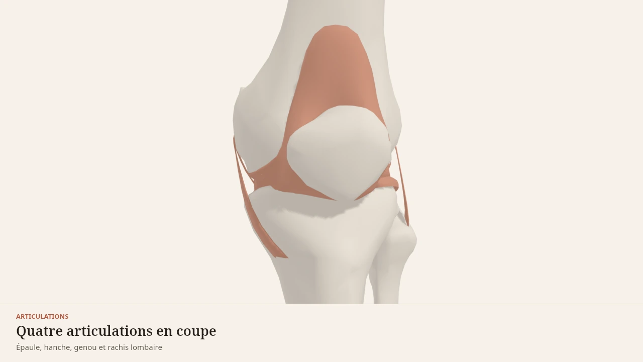
- 3/6Two poorly matched surfaces — the rounded condyles of the femur on an almost flat tibial plateau. The menisci fill the gap, the cruciates hold the tibia front to back, the collaterals hold it side to side. Remove any one of these parts and the load shifts onto the others.
- 4/6Five vertebrae, five discs, and a set of ligaments running the whole height. Each level moves only a few degrees; it is their sum that gives the trunk its range. The longitudinal ligaments limit flexion and extension, the ligamenta flava close the back of the canal.
- 5/6The elbow does two things inside one capsule. The humerus and ulna form a hinge that flexes and extends in a single plane; the head of the radius pivots in place inside a ligamentous ring, which turns the palm over. The two collateral ligaments hold it all from the sides.
- 6/6The talus is wedged between the two malleoli like a tenon in a mortise. On the inner side, a thick fan-shaped ligament; on the outer side, three markedly thinner bands. That asymmetry is no detail: it explains which way the ankle most often gives.
Model licence · Z-Anatomy et BodyParts3D — CC-BY-SA 4.0
What the model simplifies
Limit
A reference anatomy
The cartilage covering the femoral head and the floor of the socket is not represented: contact looks more direct than it is. The synovial fluid and the neighbouring bursae are also absent.
Above all, the model shows one socket orientation and one neck angle, those of the anatomical source. These two parameters are among the most variable in the skeleton, and it is precisely that variation which explains differences in range between people.
The hip is extended by the glutes and the hamstrings, flexed by the iliopsoas, and controlled sideways by the hip adductors. It works in the hip hinge, the squat and the lunge.
Sources
Main sources
- Standring S (2020). Gray's Anatomy: The Anatomical Basis of Clinical Practice, 42nd edition. Elsevier.https://shop.elsevier.com/books/grays-anatomy/standring/978-0-7020-7705-0
- Neumann DA (2016). Kinesiology of the Musculoskeletal System: Foundations for Rehabilitation, 3rd edition. Elsevier.https://shop.elsevier.com/books/kinesiology-of-the-musculoskeletal-system/neumann/978-0-323-28753-1
Put it into practice in Shapier
See the glute bridge exercise
Open the glute bridge sheet in Shapier, a hip extension exercise that targets the glutes.
See the glute bridge exercise
Body Lab explains; Shapier lets you act and track.
Check my understanding
Why does the hip move less than the shoulder?
Because it favours stability to carry the body, where the shoulder favours range to aim a hand in space
Because its ligaments are abnormally stiff
Because its muscles are weaker than the shoulder's
What makes standing cheap?
The tension of the three capsular ligaments, which tighten in extension like a rope being twisted
A continuous contraction of the glutes
The acetabular labrum, which jams the femoral head in standing
Two people perform the same squat and do not reach the same depth. What explanation does the page give?
Their bony limit is not in the same place, because socket orientation and femoral neck angle vary between people
One of them necessarily lacks flexibility and should stretch
One of them has ligaments that are too short, which limits the descent
Choose an answer
Read next
- The kneeTwo poorly matched surfaces, an almost flat plateau under rounded condyles. Menisci, cruciate and collateral ligaments make up for that lack of fit.With a 3D scene
- GlutesThe three gluteal muscles cover the back and the side of the pelvis. The gluteus maximus extends the hip, while the gluteus medius and minimus steady the pelvis as soon as the body is supported on one leg.With a 3D scene
- Hip hingeThe hip hinge concentrates the movement at the hip: the pelvis tilts over the femoral heads, the torso leans forward, and the knees stay only slightly bent. This page describes its phases, its joints, its muscles and the confusions that surround it.With a 3D scene
Available offline
Offline download is available in the mobile app.
Trust and method
Author
Editorial review
Thanh Chau
Published on
August 2, 2026
Reviewed on August 2, 2026
Next review due August 2, 2027
Limits of this page
- The articular cartilage is not represented: contact therefore looks more direct than it is.
- The orientation of the socket and the angle of the femoral neck vary noticeably between people, and that variation changes the range reachable. The model shows a reference arrangement, not an individual build.
- This page describes an anatomy. Hip or groin pain, stiffness that settles in, or a loss of range belong to a health professional: no page can attribute them to a structure.
Educational content. Body Lab does not diagnose and does not replace professional advice.
How we work