Five levels between chest and pelvis
The lumbar spine occupies the low back, between the last thoracic vertebra and the sacrum. Its five vertebrae are the most massive of the mobile spine — their bodies are wide and thick, which matches the load they carry.
Each vertebra articulates with its neighbour through three linkages: the intervertebral disc in front, and two posterior joints between the bony arches. It is the latter that, by the orientation of their surfaces, allow mainly flexion and extension, and very little rotation.
Motion segment
The unit formed by two neighbouring vertebrae, the disc separating them and the ligaments joining them. It is the smallest part of the spine that shows the mechanical properties of the whole.
functional unit
The intervertebral disc
Intervertebral disc
A fibrocartilaginous cushion between two vertebral bodies. It pairs a tough fibrous ring, arranged in crossing oblique layers, with a more deformable, water-rich central core.
disc
The disc does two things at once. It transmits load from one vertebra to the next, spreading it across the whole surface of the vertebral body. And it allows movement: the core deforms, the ring lets itself be compressed on one side and stretched on the other.
That dual function explains its make-up. A tissue too rigid would not let movement through; a tissue too soft would not transmit the load.
Certainty level · Established
The intervertebral disc transmits load between two vertebral bodies while allowing small-range movement.
Descriptive anatomy and kinesiology describe the same structure and the same function. What varies between studies is the range attributed to each level, which depends on the measurement method and the population studied.
Standring S (2020) · Neumann DA (2016)
The ligaments
A set of ligaments runs the whole height of the spine, and does not stop at the lumbar segment.
- The anterior longitudinal ligament runs down the front of the vertebral bodies. It limits extension.
- The posterior longitudinal ligament, narrower, runs along their back surface inside the canal. It limits flexion.
- The ligamenta flava close the back of the canal between successive arches. They are elastic, which lets them shorten without buckling into the canal.
- The interspinous and supraspinous ligaments join the posterior spines and limit their separation in flexion.
At the base, the iliolumbar ligament attaches the lowest lumbar vertebrae to the pelvis and limits movement at the lumbosacral junction.
Little movement per level, a lot in total
What each structure limits
Structure
Movement limited
Position where it tightens
Anterior longitudinal ligament
Extension
Arched back
Posterior longitudinal ligament
Flexion
Rounded back
Ligamenta flava
Flexion
Rounded back
Interspinous and supraspinous
Flexion
Rounded back
Disc
None on its own
It spreads load rather than limiting
A lumbar level moves only a few degrees. The trunk's apparent range comes from adding the five levels together, and above all from the hip hinge — the pelvis tips while the spine moves little.
Certainty level · Probable
The muscles framing the spine take part in transmitting force between the limbs, and their endurance is loaded in holding positions.
The review distinguishes endurance from maximal strength and describes that transmission role. It covers core training in general; turning those observations into conduct for a given person goes beyond what it establishes, and this page proposes none.
McGill SM (2010)
Seeing the lumbar spine
The lumbar spine, vertebrae and discs
The scene shows the five lumbar vertebrae and the sacrum from the side, with the five discs highlighted between them and the ligaments of the spine crossing the view from top to bottom. Without the 3D, the picture is a stack of blocks separated by cushions, held by straps running the whole height.
Current step
The lumbar spine
Five vertebrae, five discs, and a set of ligaments running the whole height. Each level moves only a few degrees; it is their sum that gives the trunk its range. The longitudinal ligaments limit flexion and extension, the ligamenta flava close the back of the canal.
Scene description
A three-dimensional model of four joints from the left side of the body, presented one after another on a paper background. The shoulder shows the shoulder blade, collarbone and upper humerus in bone tones, with the joint capsule, the glenoid labrum and the glenohumeral ligaments in olive grey. The hip shows the hip bone and femur, the capsule wrapping the neck, the acetabular labrum and the three ligaments wound around it. The knee shows the femur, tibia, fibula and kneecap, with the two menisci resting on the tibial plateau, the cruciate ligaments at the centre and the collaterals on either side. The lumbar spine, seen from the side, shows the five vertebrae and the sacrum separated by five discs, with the longitudinal ligaments running in front of and behind the vertebral bodies. The joint tissues are highlighted at each step, the bones staying in the background.
Visible structures
- Glenoid labrumA fibrocartilaginous rim fixed around the edge of the glenoid cavity. It deepens the socket and increases the contact area with the head of the humerus, whose diameter far exceeds that of the cavity.
- Ligaments of the hipThree bands wound around the capsule — iliofemoral, pubofemoral, ischiofemoral. They tighten as the hip extends, which allows standing without continuous muscular effort.
- Cruciate ligamentsTwo ligaments stretched across the centre of the knee, crossing one in front of the other. They limit the tibia sliding under the femur — forwards for the anterior, backwards for the posterior.
- Intervertebral discsFive discs separate the lumbar vertebrae and the sacrum. Each pairs a tough fibrous ring with a more deformable core, which spreads load and allows slight movement between two neighbouring vertebrae.
- Ligaments of the elbowTwo collateral ligaments hold the elbow from the sides, and a ring encircles the head of the radius so it can pivot without leaving its place. The elbow thus combines a hinge and a pivot.
- Lateral ligaments of the ankleThree slender bands link the fibula to the talus and the heel bone. They are markedly thinner than their medial counterparts, which is why the ankle is more easily forced inwards.
Guided steps
- 1/6The head of the humerus is far wider than the socket that receives it. The glenoid labrum deepens the rim, the capsule wraps the whole, and three glenohumeral ligaments reinforce it in front. That is the price of mobility: little bone to hold, a great deal of soft tissue.
- 2/6Unlike the shoulder, the femoral head sits deep in the socket, which the acetabular labrum extends further. The three ligaments wind around the neck and tighten in extension: standing, they carry part of the work the muscles no longer have to supply.
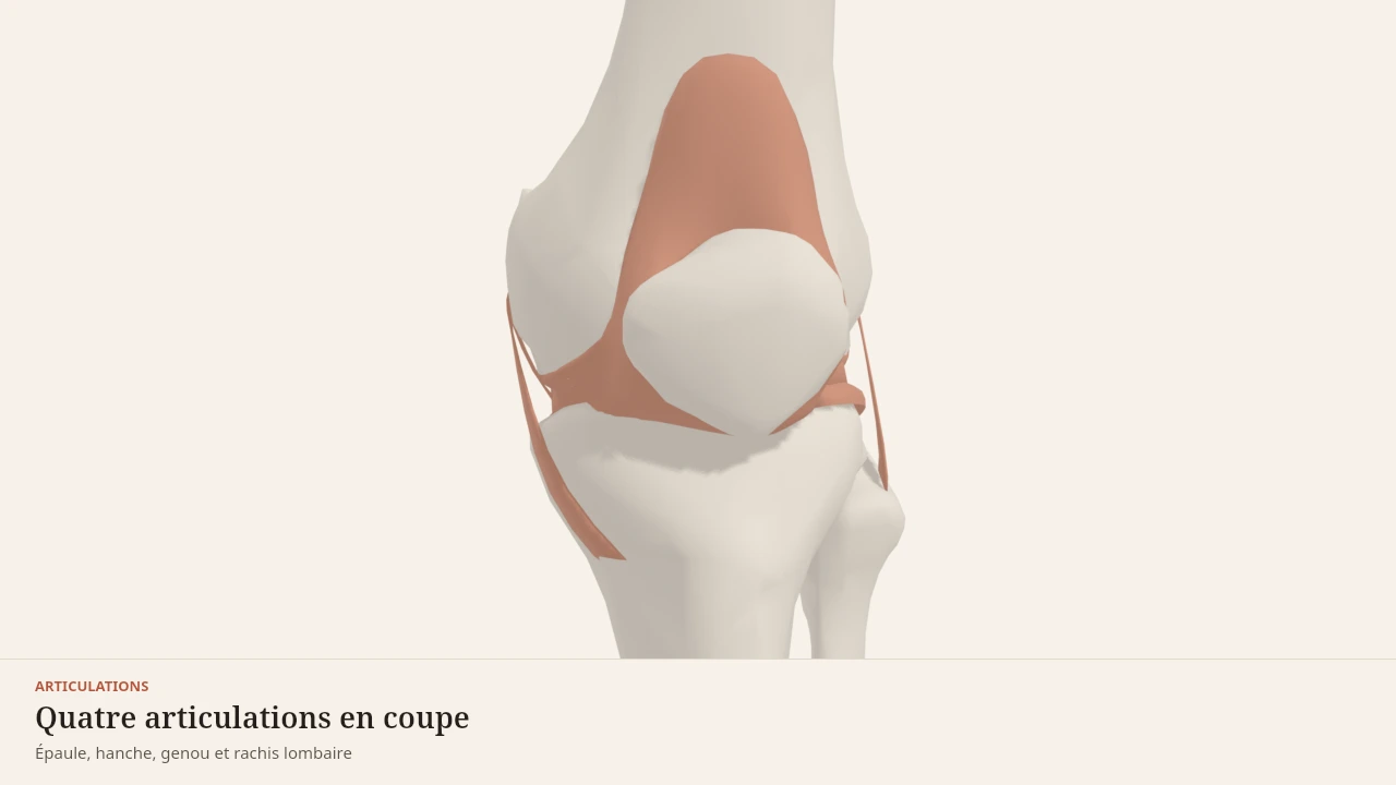
- 3/6Two poorly matched surfaces — the rounded condyles of the femur on an almost flat tibial plateau. The menisci fill the gap, the cruciates hold the tibia front to back, the collaterals hold it side to side. Remove any one of these parts and the load shifts onto the others.
- 4/6Five vertebrae, five discs, and a set of ligaments running the whole height. Each level moves only a few degrees; it is their sum that gives the trunk its range. The longitudinal ligaments limit flexion and extension, the ligamenta flava close the back of the canal.
- 5/6The elbow does two things inside one capsule. The humerus and ulna form a hinge that flexes and extends in a single plane; the head of the radius pivots in place inside a ligamentous ring, which turns the palm over. The two collateral ligaments hold it all from the sides.
- 6/6The talus is wedged between the two malleoli like a tenon in a mortise. On the inner side, a thick fan-shaped ligament; on the outer side, three markedly thinner bands. That asymmetry is no detail: it explains which way the ankle most often gives.
Model licence · Z-Anatomy et BodyParts3D — CC-BY-SA 4.0
What the model simplifies
Limit
What the image is missing
The disc is shown as a single block. Its internal structure — the fibrous ring in crossing oblique layers around the core — does not appear, although it explains most of its mechanical behaviour.
The spinal cord, the nerve roots emerging between vertebrae and the posterior joints are not depicted. The visible ligaments in fact extend beyond the lumbar segment: they are structures that run the whole spine, and the model shows them as they are rather than cutting them arbitrarily.
A subject that calls for care
Caution
What this page does not say
Back pain is one of the most frequent reasons for consultation, and its causes are many. Describing the anatomy of a disc in no way allows a pain to be attributed to that structure, and imaging is not enough either: variations are seen in people who have no pain at all.
Body Lab proposes here no exercise, no posture and no lifting rule. Pain radiating into the leg, loss of strength, altered sensation or bladder trouble require medical advice without delay.
The lumbar spine is framed by the erector spinae behind and by the abdominal wall in front. The iliopsoas attaches directly to its vertebrae. It is loaded in the hip hinge, the squat and the loaded carry.
Sources
Main sources
- Standring S (2020). Gray's Anatomy: The Anatomical Basis of Clinical Practice, 42nd edition. Elsevier.https://shop.elsevier.com/books/grays-anatomy/standring/978-0-7020-7705-0
- Neumann DA (2016). Kinesiology of the Musculoskeletal System: Foundations for Rehabilitation, 3rd edition. Elsevier.https://shop.elsevier.com/books/kinesiology-of-the-musculoskeletal-system/neumann/978-0-323-28753-1
- McGill SM (2010). Core training: evidence translating to better performance and injury prevention. Strength and Conditioning Journal.DOI : 10.1519/ssc.0b013e3181df4521
Put it into practice in Shapier
Plan deload weeks and recovery
Scheduling a lighter week and spacing out recovery time happens in Shapier.
Plan deload weeks and recovery
Body Lab explains; Shapier lets you act and track.
Check my understanding
Where does most of the trunk's apparent range come from?
From adding the five lumbar levels together, and above all from the hip hinge
From a large motion at each lumbar level
From rotation between the lumbar vertebrae
What does the intervertebral disc do?
It transmits load while spreading it and allows small-range movement
It limits trunk flexion
It limits back extension
Is a disc abnormality seen on imaging enough to explain a back pain?
No, because variations are seen in people who have no pain at all
Yes, the image lets you trace the pain back to the disc
Yes, especially if the pain radiates into the leg
Choose an answer
Read next
- Erector spinaeThe erector spinae form three muscular columns running along the spine. They straighten it, control its forward bending and hold the trunk during hip hinge movements.With a 3D scene
- The hipA sphere set deep in a socket, locked by three ligaments that tighten in extension. That is what makes standing possible without continuous muscular effort.With a 3D scene
- IliopsoasThe iliopsoas is the only muscle that links the lumbar spine directly to the femur. It flexes the hip and acts on the spine, and it is never visible from outside.With a 3D scene
Available offline
Offline download is available in the mobile app.
Trust and method
Author
Editorial review
Thanh Chau
Published on
August 2, 2026
Reviewed on August 2, 2026
Next review due August 2, 2027
Limits of this page
- The disc is shown as a single block: its internal structure — a layered fibrous ring around a more deformable core — does not appear.
- The spinal cord, the nerve roots and the posterior joints between vertebrae are not depicted, although they are central to the mechanics of the segment.
- This page describes an anatomy. Back pain is a frequent reason for consultation with many possible causes: no page can attribute it to a structure, and nothing here is a diagnosis.
- Pain radiating into the leg, loss of strength, altered sensation or bladder trouble require medical advice without delay.
Educational content. Body Lab does not diagnose and does not replace professional advice.
How we work