Three joints under one capsule
The elbow passes for a simple hinge. It is one, but not only that: three linkages share the same joint capsule, and two of them produce movement in different planes.
- The humeroulnar joint is the hinge proper. The trochlea of the humerus fits into the notch of the ulna, and the whole thing flexes and extends in a single plane.
- The humeroradial joint brings the head of the radius against the capitulum of the humerus. It accompanies flexion and allows rotation.
- The proximal radioulnar joint is a pivot: the head of the radius turns on itself against the ulna.
Hinge joint
A joint whose shape allows movement in one plane only, like a door hinge. Its stability comes largely from the fit of the bones, not from ligaments alone.
trochlear joint
The annular ligament, a ring that lets it turn
This is the elbow's most distinctive part. The annular ligament encircles the head of the radius like a band and attaches to the ulna on either side. It holds the bone without immobilising it: the radial head turns inside the ring.
That arrangement makes pronation and supination possible — the movement that rolls the palm over. The biceps brachii is its main driver, and the forearm muscles complete it in both directions.
Certainty level · Established
Forearm rotation occurs by the radius pivoting around the ulna, held at the top by the annular ligament.
Descriptive anatomy and kinesiology describe the same mechanism. What varies between studies is how the range is shared with the distal radioulnar joint at the wrist, which takes part in the same movement.
Standring S (2020) · Neumann DA (2016)
The collaterals, and the epicondyles
Two ligaments hold the elbow from the sides: the ulnar collateral inside, the radial collateral outside. They attach respectively to the medial and lateral epicondyles of the humerus — the two palpable prominences either side of the elbow.
Those same epicondyles serve as the origin of the forearm muscles: the flexors from the medial, the extensors from the lateral. A ligament and a muscle group therefore pull on the same point of bone, which is why the region comes up so often in discussions about the elbow.
What each linkage allows
Linkage
Movement
What holds it
Humeroulnar
Flexion and extension
The fit of the bones, first
Humeroradial
Accompanies both
The capsule and the collaterals
Proximal radioulnar
Pronation and supination
The annular ligament
Seeing the elbow
The elbow, bones and joint tissues
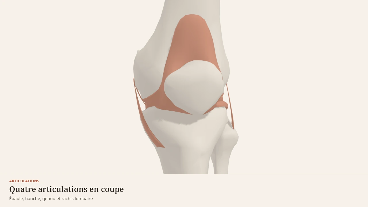
The scene shows the humerus, radius and ulna, with the capsule and ligaments highlighted. The interosseous membrane, linking the two forearm bones along their whole length, crosses the view without being its subject. Without the 3D, the picture fits in one sentence: a hinge on one side, a band on the other, inside the same wrapping.
Current step
The elbow
The elbow does two things inside one capsule. The humerus and ulna form a hinge that flexes and extends in a single plane; the head of the radius pivots in place inside a ligamentous ring, which turns the palm over. The two collateral ligaments hold it all from the sides.
Scene description
A three-dimensional model of four joints from the left side of the body, presented one after another on a paper background. The shoulder shows the shoulder blade, collarbone and upper humerus in bone tones, with the joint capsule, the glenoid labrum and the glenohumeral ligaments in olive grey. The hip shows the hip bone and femur, the capsule wrapping the neck, the acetabular labrum and the three ligaments wound around it. The knee shows the femur, tibia, fibula and kneecap, with the two menisci resting on the tibial plateau, the cruciate ligaments at the centre and the collaterals on either side. The lumbar spine, seen from the side, shows the five vertebrae and the sacrum separated by five discs, with the longitudinal ligaments running in front of and behind the vertebral bodies. The joint tissues are highlighted at each step, the bones staying in the background.
Visible structures
- Glenoid labrumA fibrocartilaginous rim fixed around the edge of the glenoid cavity. It deepens the socket and increases the contact area with the head of the humerus, whose diameter far exceeds that of the cavity.
- Ligaments of the hipThree bands wound around the capsule — iliofemoral, pubofemoral, ischiofemoral. They tighten as the hip extends, which allows standing without continuous muscular effort.
- Cruciate ligamentsTwo ligaments stretched across the centre of the knee, crossing one in front of the other. They limit the tibia sliding under the femur — forwards for the anterior, backwards for the posterior.
- Intervertebral discsFive discs separate the lumbar vertebrae and the sacrum. Each pairs a tough fibrous ring with a more deformable core, which spreads load and allows slight movement between two neighbouring vertebrae.
- Ligaments of the elbowTwo collateral ligaments hold the elbow from the sides, and a ring encircles the head of the radius so it can pivot without leaving its place. The elbow thus combines a hinge and a pivot.
- Lateral ligaments of the ankleThree slender bands link the fibula to the talus and the heel bone. They are markedly thinner than their medial counterparts, which is why the ankle is more easily forced inwards.
Guided steps
- 1/6The head of the humerus is far wider than the socket that receives it. The glenoid labrum deepens the rim, the capsule wraps the whole, and three glenohumeral ligaments reinforce it in front. That is the price of mobility: little bone to hold, a great deal of soft tissue.
- 2/6Unlike the shoulder, the femoral head sits deep in the socket, which the acetabular labrum extends further. The three ligaments wind around the neck and tighten in extension: standing, they carry part of the work the muscles no longer have to supply.
- 3/6Two poorly matched surfaces — the rounded condyles of the femur on an almost flat tibial plateau. The menisci fill the gap, the cruciates hold the tibia front to back, the collaterals hold it side to side. Remove any one of these parts and the load shifts onto the others.
- 4/6Five vertebrae, five discs, and a set of ligaments running the whole height. Each level moves only a few degrees; it is their sum that gives the trunk its range. The longitudinal ligaments limit flexion and extension, the ligamenta flava close the back of the canal.
- 5/6The elbow does two things inside one capsule. The humerus and ulna form a hinge that flexes and extends in a single plane; the head of the radius pivots in place inside a ligamentous ring, which turns the palm over. The two collateral ligaments hold it all from the sides.
- 6/6The talus is wedged between the two malleoli like a tenon in a mortise. On the inner side, a thick fan-shaped ligament; on the outer side, three markedly thinner bands. That asymmetry is no detail: it explains which way the ankle most often gives.
Model licence · Z-Anatomy et BodyParts3D — CC-BY-SA 4.0
What the model simplifies
Limit
What the image is missing
The cartilage is not represented, nor are the bursae. The olecranon bursa, at the point of the elbow, accounts for part of the swelling seen in the region.
Forearm rotation does not happen at the elbow alone: the distal radioulnar joint at the wrist takes part and does not appear here. The model shows the left side, in a reference anatomy.
The elbow is flexed by the biceps brachii and brachialis, extended by the triceps brachii. The forearm muscles attach to it via the epicondyles and transmit force to the hand. It works in every pull and in the loaded carry.
Sources
Main sources
- Standring S (2020). Gray's Anatomy: The Anatomical Basis of Clinical Practice, 42nd edition. Elsevier.https://shop.elsevier.com/books/grays-anatomy/standring/978-0-7020-7705-0
- Neumann DA (2016). Kinesiology of the Musculoskeletal System: Foundations for Rehabilitation, 3rd edition. Elsevier.https://shop.elsevier.com/books/kinesiology-of-the-musculoskeletal-system/neumann/978-0-323-28753-1
Put it into practice in Shapier
See the biceps curl exercise
Open the biceps curl page in Shapier, the exercise that works the biceps brachii.
See the biceps curl exercise
Body Lab explains; Shapier lets you act and track.
Check my understanding
The elbow is a simple hinge: true or false?
True only in part: it is a hinge, but three linkages share the same capsule, and two of them produce movement in different planes.
True: it only flexes and extends the arm.
False: it does not flex, it only pivots.
What does the annular ligament do?
It holds the head of the radius against the ulna without immobilising it, so the bone can turn.
It immobilises the radius to stabilise the elbow.
It connects the radius to the humerus on either side.
Where does forearm rotation actually happen?
At the elbow and at the wrist: the distal radioulnar joint takes part in the same movement as the proximal one.
Only at the elbow, in the proximal radioulnar joint.
Only at the wrist, the elbow merely flexing.
Choose an answer
Read next
- The ankleThe talus is wedged between two malleoli like a tenon in a mortise. The ligaments holding it are not symmetrical, and that asymmetry explains a great deal.With a 3D scene
- Forearm musclesThe forearm muscles drive the wrist and fingers from a distance, through long tendons. They carry grip strength, which often gives out before the large muscles do.With a 3D scene
- The shoulderThe most mobile joint in the body, and the one that holds least by its shape. A wide head resting on a shallow socket, retained by soft tissue.With a 3D scene
Available offline
Offline download is available in the mobile app.
Trust and method
Author
Editorial review
Thanh Chau
Published on
August 2, 2026
Reviewed on August 2, 2026
Next review due August 2, 2027
Limits of this page
- The cartilage is not represented, nor are the bursae: the olecranon bursa, at the point of the elbow, accounts for part of the swelling seen in the region.
- The distal radioulnar joint at the wrist takes part in the same rotation but is not depicted: pronation does not happen at the elbow alone.
- This page describes an anatomy. Epicondyle pain, stiffness that settles in, or loss of rotation belong to a health professional: no page can attribute them to a structure.
Educational content. Body Lab does not diagnose and does not replace professional advice.
How we work