Not a frame
Body Lab's scenes show bones as pale, smooth volumes, placed there to carry the rest. That is convenient and wrong on one essential point: a bone is living tissue, threaded with vessels and nerves, taken apart and rebuilt without pause.
Two arrangements, one material
Bone is not solid. It combines two arrangements of the same material, distributed according to the stresses it takes.
- Compact bone forms the outer shell, dense and strong. It is thickest at the middle of long bones, where bending is greatest.
- Spongy bone fills the inside of the ends. It is made of trabeculae — thin bony walls — oriented along the forces crossing the region.
Trabecula
A thin wall of spongy bone inside the ends of a bone. The orientation of trabeculae follows the lines of stress: it differs from bone to bone, and changes if the loading changes.
bony strut
That orientation is visible on a section through the femoral neck: the trabeculae draw arcs matching the compression and tension forces crossing the hip. Bone does not merely resist load, it arranges itself according to it.
A permanent worksite
Two populations of cells work in opposite directions, without ever stopping.
Two cells, two roles
Cell
What it does
Effect
Osteoclast
Dissolves the bone matrix
Removes material
Osteoblast
Lays down new matrix
Adds material
Osteocyte
Cell walled into the bone
Senses stress and signals
This cycle of removal and rebuilding is called remodelling. It is continuous, including in adults, and each year it touches a notable share of the skeleton. A bone is therefore not the same material from one year to the next, even if its shape does not change.
Certainty level · Established
Bone tissue is continuously remodelled, and the orientation of its trabeculae matches the mechanical stresses it takes.
Descriptive anatomy and kinesiology describe the same organisation and the same cellular mechanism. The correspondence between trabecular orientation and lines of stress is observable on section. Turnover rates vary with age, bone site and hormonal state.
Standring S (2020) · Neumann DA (2016)
What adapts, and how fast
Like the tendon, bone adapts to load, and like it, more slowly than muscle. The signal it responds to is not repetition but deformation: it is unusual stress, in size or in direction, that triggers remodelling.
That relative slowness has the same practical consequence as for tendon: the capacity to produce force can improve before the load-bearing tissue has finished adjusting. Body Lab draws no training rule from this — it is an observation about structure.
Key point
Three tissues, three rhythms
Muscle turns over fast and signals its fatigue. Tendon adapts more slowly and warns late. Bone is slower still, and barely warns at all.
That is why the three do not read on the same horizon: what shows in weeks for one is measured in months for the others.
Seeing a bone
The bones of the joints
The scene shows the hip bone and the femur in bone tones. What you see is the surface: the internal structure, the marrow, the periosteum and the vessels do not appear. Without the 3D, the idea fits in one sentence: what looks solid is in fact a dense shell around a network of oriented struts.
Current step
The hip
Unlike the shoulder, the femoral head sits deep in the socket, which the acetabular labrum extends further. The three ligaments wind around the neck and tighten in extension: standing, they carry part of the work the muscles no longer have to supply.
Scene description
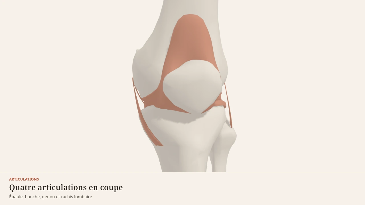
A three-dimensional model of four joints from the left side of the body, presented one after another on a paper background. The shoulder shows the shoulder blade, collarbone and upper humerus in bone tones, with the joint capsule, the glenoid labrum and the glenohumeral ligaments in olive grey. The hip shows the hip bone and femur, the capsule wrapping the neck, the acetabular labrum and the three ligaments wound around it. The knee shows the femur, tibia, fibula and kneecap, with the two menisci resting on the tibial plateau, the cruciate ligaments at the centre and the collaterals on either side. The lumbar spine, seen from the side, shows the five vertebrae and the sacrum separated by five discs, with the longitudinal ligaments running in front of and behind the vertebral bodies. The joint tissues are highlighted at each step, the bones staying in the background.
Visible structures
- Glenoid labrumA fibrocartilaginous rim fixed around the edge of the glenoid cavity. It deepens the socket and increases the contact area with the head of the humerus, whose diameter far exceeds that of the cavity.
- Ligaments of the hipThree bands wound around the capsule — iliofemoral, pubofemoral, ischiofemoral. They tighten as the hip extends, which allows standing without continuous muscular effort.
- Cruciate ligamentsTwo ligaments stretched across the centre of the knee, crossing one in front of the other. They limit the tibia sliding under the femur — forwards for the anterior, backwards for the posterior.
- Intervertebral discsFive discs separate the lumbar vertebrae and the sacrum. Each pairs a tough fibrous ring with a more deformable core, which spreads load and allows slight movement between two neighbouring vertebrae.
- Ligaments of the elbowTwo collateral ligaments hold the elbow from the sides, and a ring encircles the head of the radius so it can pivot without leaving its place. The elbow thus combines a hinge and a pivot.
- Lateral ligaments of the ankleThree slender bands link the fibula to the talus and the heel bone. They are markedly thinner than their medial counterparts, which is why the ankle is more easily forced inwards.
Guided steps
- 1/6The head of the humerus is far wider than the socket that receives it. The glenoid labrum deepens the rim, the capsule wraps the whole, and three glenohumeral ligaments reinforce it in front. That is the price of mobility: little bone to hold, a great deal of soft tissue.
- 2/6Unlike the shoulder, the femoral head sits deep in the socket, which the acetabular labrum extends further. The three ligaments wind around the neck and tighten in extension: standing, they carry part of the work the muscles no longer have to supply.
- 3/6Two poorly matched surfaces — the rounded condyles of the femur on an almost flat tibial plateau. The menisci fill the gap, the cruciates hold the tibia front to back, the collaterals hold it side to side. Remove any one of these parts and the load shifts onto the others.
- 4/6Five vertebrae, five discs, and a set of ligaments running the whole height. Each level moves only a few degrees; it is their sum that gives the trunk its range. The longitudinal ligaments limit flexion and extension, the ligamenta flava close the back of the canal.
- 5/6The elbow does two things inside one capsule. The humerus and ulna form a hinge that flexes and extends in a single plane; the head of the radius pivots in place inside a ligamentous ring, which turns the palm over. The two collateral ligaments hold it all from the sides.
- 6/6The talus is wedged between the two malleoli like a tenon in a mortise. On the inner side, a thick fan-shaped ligament; on the outer side, three markedly thinner bands. That asymmetry is no detail: it explains which way the ankle most often gives.
Model licence · Z-Anatomy et BodyParts3D — CC-BY-SA 4.0
What the model simplifies
Limit
A surface, not a tissue
The models render bone as a smooth, uniform shell. Three things are missing, and they matter.
The periosteum, the membrane wrapping the bone and carrying its vessels and nerve endings. The internal structure, which distinguishes a spongy end from a compact shaft. And the marrow, housed in the cavities, whose role goes beyond the scope of these pages.
A model bone is a shape. A real bone is a tissue with a mechanical history.
What this page does not do
Body Lab describes mechanisms. This page assesses no bone density, does not say how to gain or preserve any, and replaces no examination.
Localised bone pain, a fracture from a minor impact, or unexplained loss of height belong to a health professional.
Sources
Main sources
- Standring S (2020). Gray's Anatomy: The Anatomical Basis of Clinical Practice, 42nd edition. Elsevier.https://shop.elsevier.com/books/grays-anatomy/standring/978-0-7020-7705-0
- Neumann DA (2016). Kinesiology of the Musculoskeletal System: Foundations for Rehabilitation, 3rd edition. Elsevier.https://shop.elsevier.com/books/kinesiology-of-the-musculoskeletal-system/neumann/978-0-323-28753-1
Put it into practice in Shapier
Practise the squat, a load-bearing exercise
Reviewing squat technique belongs in Shapier; Body Lab only explains how bone remodels under load.
Practise the squat, a load-bearing exercise
Body Lab explains; Shapier lets you act and track.
Check my understanding
What separates a real bone from an inert frame?
It is living tissue, threaded with vessels and nerves, taken apart and rebuilt without pause.
It is a finished material once growth is over.
It is a solid beam, dense through its whole thickness.
According to this page, what signal triggers bone remodelling?
Deformation, meaning stress that is unusual in size or in direction.
Repeating the same movement, through sheer accumulation.
Fatigue signalled by the neighbouring muscle.
How do the three tissues rank by speed of adaptation?
Muscle adapts fast, tendon more slowly, bone more slowly still.
Tendon is the slowest of the three.
All three adapt at the same pace since they all respond to load.
Choose an answer
Read next
- CartilageThe surface two bones glide on. No vessels, no nerves, fed by movement itself — and absent from every scene, as six joint pages already point out.Described without a scene
- The tendonThe part that links a muscle to a bone. It transmits force, briefly stores it like a spring, and adapts far more slowly than the muscle it continues.With a 3D scene
- FasciaA continuous sheet of connective tissue, real and well described in anatomy. What is commonly said about it goes markedly further than what the literature establishes.Described without a scene
Available offline
Offline download is available in the mobile app.
Trust and method
Author
Editorial review
Thanh Chau
Published on
August 2, 2026
Reviewed on August 2, 2026
Next review due August 2, 2027
Limits of this page
- The scenes show smooth, uniform bone surfaces: neither the internal trabecular structure, nor the marrow, nor the periosteum appears.
- The adaptation times quoted describe a trend from bone physiology: they vary widely with age, bone site and hormonal state.
- This page describes a tissue. It assesses no bone density, does not say how to gain any, and replaces no examination.
- Localised bone pain, a fracture from a minor impact, or unexplained loss of height belong to a health professional.
Educational content. Body Lab does not diagnose and does not replace professional advice.
How we work