Every muscle contraction spends the same currency: ATP. The cell keeps very little of it in reserve, and yet the effort carries on. The video shows why, by following a single molecule through a sprint and then through the rest that follows.
The ATP–phosphocreatine system
Duration: 1:24
Chapters
Transcript
ATP is the only molecule a muscle fibre spends directly in order to contract. The store present in the muscle is very small: it has to be rebuilt continuously.
When ATP gives up a phosphate, it becomes ADP and releases energy. Phosphocreatine immediately donates a phosphate back to that ADP to reform ATP. This reaction needs neither oxygen nor delay: it is the fastest resynthesis pathway.
The phosphocreatine store supports an all-out effort for a few seconds only, then rebuilds during rest over a few minutes through aerobic metabolism. Rest duration between sets follows directly from that kinetics.
As effort lasts, glycolysis and then aerobic metabolism take over. The pathways do not work like switches: they overlap continuously, and the contribution of each depends on the intensity and duration of the effort.
What the video shows
The animation opens on a schematic muscle cell. One molecule of ATP moves into contact with the contractile filaments, gives up one of its phosphate groups, and becomes ADP. The movement happens; the currency has been spent.
The shot then widens onto the reserve available. It is visibly tiny: the video draws it as a handful of molecules, used up within a few contractions. This is the central point of the sequence — the cell does not store energy as ATP, it manufactures it continuously.
Then comes phosphocreatine. A second molecule approaches the ADP, hands back a phosphate group, and rebuilds the ATP straight away. No intermediate step, no oxygen consumed: it is the fastest route the muscle has. The video repeats that transfer several times, until the phosphocreatine store falls in its turn.
The closing shot overlays three curves in different colours, which rise and fall together rather than taking over from one another. The video ends on the rest period: at rest, the transfer runs the other way and phosphocreatine is rebuilt from the ATP produced by aerobic metabolism.
Three routes, one product
Route
Speed of supply
Total capacity
Oxygen required
ATP–phosphocreatine
The fastest
The smallest
No
Glycolysis
Fast
Intermediate
No
Aerobic metabolism
The slowest to ramp up
The largest
Yes
Why the recharge matters as much as the spending
The least intuitive part of the video is the ending, not the opening. Phosphocreatine is not a fuel that gets burned: it is a rechargeable buffer. How far it is rebuilt during recovery periods shapes the quality of the next effort, which explains why the length of the rest between sets changes the feel of a session of short efforts so much.
Certainty level · Established
Phosphocreatine acts as a buffer store that regenerates ATP almost instantly, then rebuilds itself during recovery.
Reviews of how the metabolic routes interact describe this phosphate group transfer as the fastest source of ATP in skeletal muscle, and its resynthesis as a process that depends on oxidative metabolism during rest. The rates measured vary with fibre type, with the muscle studied and with the method used; the data most often come from maximal efforts performed in a laboratory.
Baker JS, McCormick MC, Robergs RA (2010)
Pathways that overlap rather than take turns
The classic picture of three pathways handing over to one another is convenient, but the video corrects it explicitly with its three overlapping curves.
Certainty level · Established
During a maximal effort, the anaerobic and aerobic routes contribute at the same time, and the aerobic share becomes dominant earlier than the classic account suggests.
A review of energy system interaction reports that, during continuous maximal exercise, the anaerobic and aerobic contributions even out within a few tens of seconds, with values that vary with the protocol, the ergometer and the participants' level. The studies gathered involve healthy adults and continuous efforts: they do not directly describe a resistance session broken up by rest periods.
Gastin PB (2001)
The linked scene
The ATP–phosphocreatine cycle
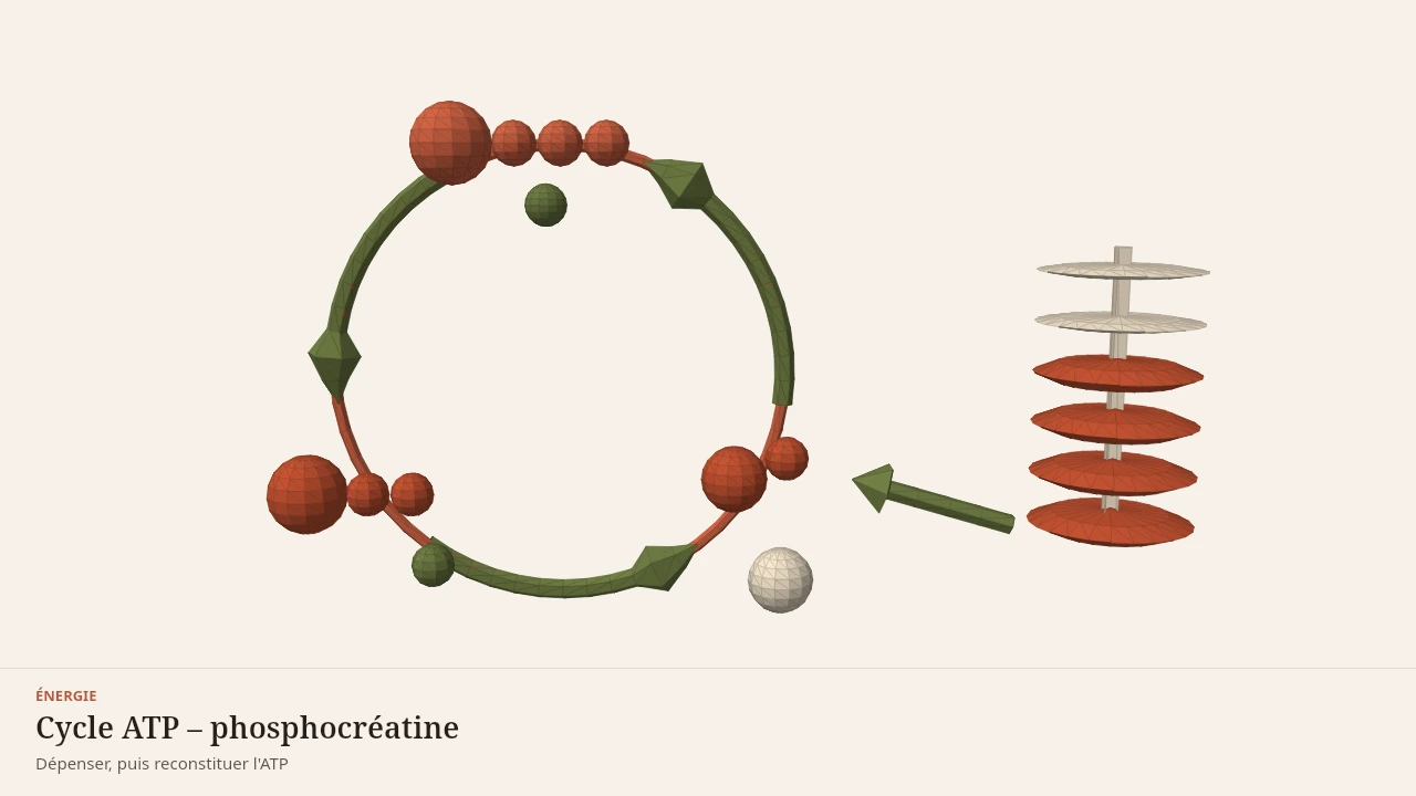
The scene takes the phosphate group transfer shown in the video and lets you explore it step by step, in both directions: spending during the contraction, recharging during the rest. Every step is described in text below the scene and stays understandable without displaying the model.
Current step
1. A loop, not a line
Cellular energy runs in a closed loop: ATP is spent, then rebuilt. A muscle stores almost no ATP in advance; it recycles it continuously.
Scene description
Circular diagram: a pale ring carries three stations linked by arrows running anticlockwise. At the top, ATP is drawn as a large adenosine sphere followed by three small phosphate spheres; at the bottom left, ADP carries only two of them and a detached phosphate floats alongside; at the bottom right, phosphocreatine hands over its phosphate and leaves a creatine sphere behind. To the right of the circle, six discs stacked on a spindle represent the phosphocreatine store: the four thick discs at the bottom are still available, the two thin discs at the top have already been used, and an arrow links the store to the resynthesis station. A small marker travels around the ring to follow the cycle. The volumes are symbolic: neither the shape nor the size of the molecules is depicted.
Visible structures
- ATPAdenosine triphosphate carries three phosphate groups. It is the form of energy a muscle fibre uses directly in order to contract.
- ADP and free phosphateWhen ATP gives up a phosphate, ADP and a free phosphate are left behind and the energy of the bond becomes available. The cell then has to rebuild ATP.
- PhosphocreatinePhosphocreatine stored in the muscle hands its phosphate to ADP, which rebuilds ATP almost immediately. What remains is creatine.
- Phosphocreatine storeThe stack stands for a limited store: the solid discs are still available, the pale ones have already been used. It is rebuilt during recovery.
- CreatineOnce its phosphate has been handed over, creatine is what remains. It is phosphorylated again when the effort stops and energy becomes available.
Guided steps
- 1/5Cellular energy runs in a closed loop: ATP is spent, then rebuilt. A muscle stores almost no ATP in advance; it recycles it continuously.
- 2/5ATP carries three phosphates, shown here as three small spheres in a row. It is the only form of energy the contractile proteins can use directly.
- 3/5By releasing its third phosphate, ATP becomes ADP and frees the energy that powers contraction. The detached phosphate stays available inside the cell.
- 4/5Phosphocreatine transfers its phosphate to ADP: ATP is rebuilt almost instantly, without oxygen. It is the fastest route a muscle has.
- 5/5The stack of discs stands for the phosphocreatine store: it supports a very short, very intense effort, then runs down. It is rebuilt during recovery, once the other pathways take over.
Model licence · Shapier — Propriétaire — usage interne ShapierLab
Limit
What the video simplifies
Molecules are drawn as solid objects of comparable size, which they are not. Where they sit in the cell is schematic too.
The animation leaves out the detail of the mitochondria, the fate of hydrogen ions, and the exchanges between the muscle and the rest of the body during effort.
Sources for this video
- Gastin PB (2001). Energy system interaction and relative contribution during maximal exercise. Sports Medicine.DOI : 10.2165/00007256-200131100-00003
- Baker JS, McCormick MC, Robergs RA (2010). Interaction among skeletal muscle metabolic energy systems during intense exercise. Journal of Nutrition and Metabolism.DOI : 10.1155/2010/905612
Read next
- ATP and phosphocreatineWhere does the energy for the very first seconds of a maximal effort come from? This page describes the energy currency of muscle, the buffering role of phosphocreatine, and why the energy systems do not take turns the way they are usually described.With a 3D scene
- Muscle glycogen and waterWhy can the number on the scales change within two days without any change in fat mass? This page explains what muscle glycogen is, its role as a fuel and the water that accompanies it inside the muscle.With a 3D scene
- Energy and recoveryA six-step reading pathway that follows muscle energy from the second to the night: the instant recharge of ATP, the glycogen stores and their water, then the role of sleep. A closing quiz lets you check what you have taken in.Described without a scene
Put it into practice in Shapier
Read the advice on recovery weeks
Read in Shapier the advice page that explains how to place lighter weeks and rest periods within a training progression.
Read the advice on recovery weeks
Body Lab explains; Shapier lets you act and track.
Available offline
Offline download is available in the mobile app.
Trust and method
Author
Editorial review
Thanh Chau
Published on
August 2, 2026
Reviewed on August 2, 2026
Next review due August 2, 2027
Limits of this page
- The animation shows a schematic muscle cell: the proportions between molecules, organelles and distances are chosen for legibility, never drawn to scale.
- The studies cited involve maximal efforts performed in a laboratory by healthy adults; the durations observed vary with the protocol, the muscle tested and the training level.
- Splitting metabolism into three "pathways" is a teaching convention: the metabolic routes run at the same time rather than one after the other.
Educational content. Body Lab does not diagnose and does not replace professional advice.
How we work