The squat looks simple: you go down, you come back up. Seen from the inside, three joints change angle at the same time and share the work between them. The video slows the movement down and shows how that sharing happens, shot by shot.
The mechanics of the squat
Duration: 1:22
Transcript
The squat is a coordinated flexion of the hip, knee and ankle. The three joints descend and rise together, and that coordination is what makes the movement stable.
On the way down, the extensor muscles brake the movement while lengthening under tension. The knee travels forward, the hip travels back and the trunk inclines: these three adjustments go together. Achievable depth depends notably on ankle mobility and segment proportions, which vary between individuals.
On the way up, the quadriceps extend the knee while the glutes extend the hip. The spinal erectors and the abdominal wall keep the trunk rigid so force can be transmitted. The share of work between knee and hip changes with bar position.
A high-bar squat loads the knee extensors more; a low-bar squat, more inclined, shifts the demand towards the hip extensors. No variation is superior in absolute terms: they simply distribute the work differently.
What the video shows
The animation opens on a side view with the bones visible. As the descent begins, three angles close at once: the hip flexes, the knee flexes, the ankle moves into dorsiflexion — the shin tips forward over the foot. The video insists on that simultaneity: no joint works on its own.
A vertical line then appears, the line of the load. The horizontal distance between that line and each joint is the moment arm. The longer that distance, the more force the muscles crossing the joint have to produce to control it. By tilting the torso, the video shows the moment arm at the hip lengthening while the one at the knee shortens, and the other way round.
The next shot brings in the muscles. The quadriceps control the knee, the glutes and hamstrings act on the hip, the calves stabilise the ankle, the erector spinae and the abdominal wall keep the trunk braced. The ascent runs the same sequence in reverse.
The phases of the squat in the video
- 1Starting positionBefore the movementHip, knee and ankle close to extension, load lined up over the middle of the foot.
- 2DescentEccentric phaseThe three joints flex together; the muscles slow the descent while lengthening under tension.
- 3Bottom positionTransitionThe moment arms are at their longest; the direction of travel reverses with no marked pause.
- 4AscentConcentric phaseThe muscles shorten under tension and re-extend the three joints back to the starting position.
The video does not try to name a "correct" technique; it makes a trade-off visible. A more upright torso brings the load closer to the hip and further from the knee: the work shifts towards the quadriceps. A more inclined torso does the opposite and asks more of the hip.
Certainty level · Established
The squat is a multi-joint movement in which hip, knee and ankle share the mechanical demand according to body geometry and the position of the load.
Kinesiology textbooks describe the moment produced by an external force as that force multiplied by its moment arm about the joint axis. This framework explains why two people built differently spontaneously adopt different angles in the same exercise. It describes general mechanical principles and does not predict the best technique for any given person.
Neumann DA (2016)
What changes with depth
One of the most useful shots in the video overlays two depths of descent and displays how the loads at the knee evolve.
Certainty level · Probable
Compressive forces at the knee increase with the flexion angle reached during the squat.
A biomechanical analysis of the dynamic squat reports that patellofemoral and tibiofemoral compressive forces rise as the knee flexes, while the load on the anterior cruciate ligament stays low throughout the movement. Those measurements were obtained in trained lifters, in a laboratory, with controlled loads. A higher load means a greater mechanical demand, which reviews on hypertrophy identify as the trigger for adaptation; it is not in itself a sign of danger.
Escamilla RF (2001) · Schoenfeld BJ (2010)
The linked scene
Quadriceps and the squat
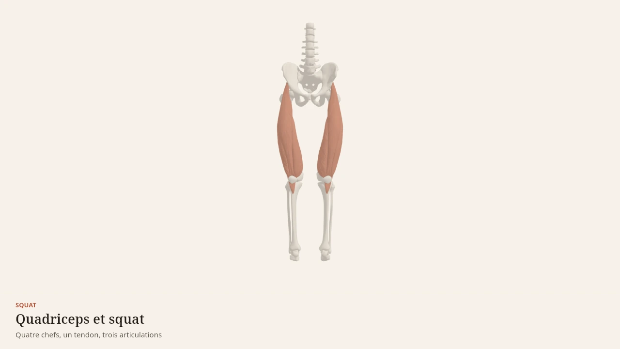
The scene takes the side view from the video and lets you vary the depth of the squat step by step, with the quadriceps isolated. Every step is described in text below the scene and stays understandable without displaying the model.
Current step
Three segments, three joints
The squat is not a thigh movement: it is a sequence. Pelvis, femur and tibia form three segments linked by the hip, the knee and the ankle, with the patella sitting in front of the knee. The whole mechanics of the movement lives in those pivots.
Scene description
A three-dimensional diagram of the lower half of the body, standing, seen from the front: pelvis, femurs, tibias and patellae are shown in bone tones, with the feet and a reference trunk in a neutral paper shade. On each thigh the four heads of the quadriceps are laid over in terracotta and stay distinct: rectus femoris in the middle, vastus lateralis on the outside, vastus medialis on the inside, and the deeper vastus intermedius against the femur. Behind, the glutes cap the pelvis and the calves fill the upper shank. A three-second animation flexes the ankle, knee and hip together: the knee travels forward over the foot, the hip moves back and down, the trunk leans, and then the movement reverses. The volumes are stylised: this is a teaching diagram, not an exact anatomical reconstruction.
Visible structures
- Rectus femorisThe only head of the quadriceps to cross two joints: it arises from the anterior inferior iliac spine of the pelvis and runs down the middle of the thigh into the quadriceps tendon. It extends the knee and flexes the hip.
- Vastus lateralisThe largest of the four heads, arising from the greater trochanter and the linea aspera on the outer side of the femur. It acts on the knee alone, extending it, and gives the thigh its outer contour.
- Vastus medialisThe inner head, arising from the linea aspera of the femur, whose lowest fibres reach the patella almost horizontally. It extends the knee and helps keep the patella tracking in line.
- Vastus intermediusThe deep head, pressed against the front of the femur and covered by the other three. Invisible from the outside, it still produces a substantial share of knee extension.
- PatellaA sesamoid bone embedded in the quadriceps tendon, which continues as the patellar tendon onto the tibial tuberosity. By holding the tendon away from the knee axis, it increases the lever arm of the quadriceps.
- GlutesArising from the iliac wing and the sacrum, inserting on the femur and the iliotibial tract. In the squat they extend the hip: the more the trunk leans forward, the larger their share of the work compared with the quadriceps.
- CalvesThe gastrocnemius and soleus, joining on the calcaneus through the Achilles tendon. In the squat they work mostly as brakes, controlling ankle dorsiflexion and therefore how far the knee travels forward.
Guided steps
- 1/5The squat is not a thigh movement: it is a sequence. Pelvis, femur and tibia form three segments linked by the hip, the knee and the ankle, with the patella sitting in front of the knee. The whole mechanics of the movement lives in those pivots.
- 2/5Rectus femoris occupies the middle of the thigh, vastus lateralis its outer border, vastus medialis its inner border, and vastus intermedius hides beneath them against the femur. All four converge on a single tendon: they pull together, but only rectus femoris also crosses the hip.
- 3/5The quadriceps tendon wraps around the patella and continues to the tibial tuberosity. By holding the tendon away from the axis of flexion, the patella lengthens the lever arm — that is what makes knee extension effective in the bottom position.
- 4/5Ankle, knee and hip flex together. The ankle lets the knee travel forward over the foot, the femur tips backwards, and the trunk leans to keep balance over the midfoot. The quadriceps first brakes the descent, then drives the way back up.
- 5/5The glutes extend the hip and take on more work the further the trunk leans; the calves restrain the ankle and steady the base. The quadriceps is the most heavily loaded engine in the squat, but it does not decide the depth reached on its own.
Model licence · Z-Anatomy et BodyParts3D — CC-BY-SA 4.0
Caution
What this page does not do
Body Lab explains a movement, never an individual situation. No technical correction, no diagnosis and no personal adjustment can be drawn from this page.
Pain that appears, persists or limits movement is a matter for a healthcare professional who can examine the person concerned.
Sources for this video
- Neumann DA (2016). Kinesiology of the Musculoskeletal System: Foundations for Rehabilitation, 3rd edition. Elsevier.https://shop.elsevier.com/books/kinesiology-of-the-musculoskeletal-system/neumann/978-0-323-28753-1
- Escamilla RF (2001). Knee biomechanics of the dynamic squat exercise. Medicine and Science in Sports and Exercise.DOI : 10.1097/00005768-200101000-00020
- Schoenfeld BJ (2010). The mechanisms of muscle hypertrophy and their application to resistance training. Journal of Strength and Conditioning Research.DOI : 10.1519/jsc.0b013e3181e840f3
Read next
- SquatThe squat is a simultaneous flexion, then extension, of the hips, knees and ankles with the feet planted on the ground. This page describes its phases, the joints involved, the muscles that contribute and the most widespread misconceptions about it.With a 3D scene
- QuadricepsThe quadriceps brings together four muscles on the front of the thigh that share a common tendon. It extends the knee, and one of its heads also crosses the hip.With a 3D scene
- Understanding muscle growthA six-step reading pathway that connects the mechanical signal produced during a set, the protein building that follows, and the volume of work accumulated over a week. A quiz at the end lets you check what you have taken in.Described without a scene
Put it into practice in Shapier
Perform the guided squat
Performing the squat step by step happens in Shapier; Body Lab only explains the biomechanics of the movement.
Perform the guided squat
Body Lab explains; Shapier lets you act and track.
Available offline
Offline download is available in the mobile app.
Trust and method
Author
Editorial review
Thanh Chau
Published on
August 2, 2026
Reviewed on August 2, 2026
Next review due August 2, 2027
Limits of this page
- The animation uses a reference skeleton with average proportions: the relative length of the segments varies from one person to the next and changes how the movement looks.
- The joint loads quoted come from laboratory analyses carried out on small groups of trained lifters, under controlled conditions.
- A high mechanical load is not the same thing as an injury: the literature cited describes forces, not an individual risk.
Educational content. Body Lab does not diagnose and does not replace professional advice.
How we work